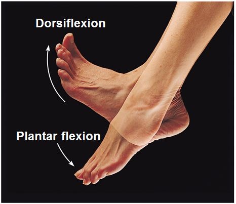
diagrams
0.0(0)
Card Sorting
1/108
Earn XP
Description and Tags
Last updated 1:22 AM on 11/4/22
Name | Mastery | Learn | Test | Matching | Spaced | Call with Kai |
|---|
No analytics yet
Send a link to your students to track their progress
109 Terms
1
New cards
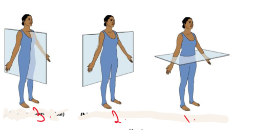
1. transverse plane
2. Frontal ( coronal) plane
3. median ( midsagittal)
2. Frontal ( coronal) plane
3. median ( midsagittal)
anatomical planes
1.
2.
3
1.
2.
3
2
New cards
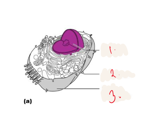
1. nucleus
2. cytoplasm
3. plasma membrane
2. cytoplasm
3. plasma membrane
Cell diagram
1.
2.
3.
1.
2.
3.
3
New cards
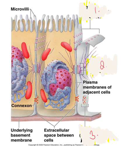
1. tight junctions
2. desmosome
3. tight junctions
2. desmosome
3. tight junctions
1.
2.
3.
2.
3.
4
New cards
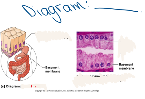
simple columnar epithelium
Made up of a single layer of tall cells that fit closely together
5
New cards
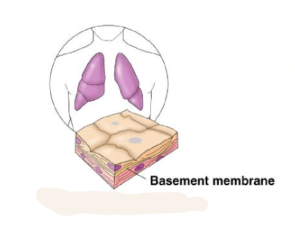
simple squamous epithelium
single layer of flattened cells
6
New cards
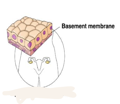
simple cuboidal
single layer of cube shaped cells
7
New cards
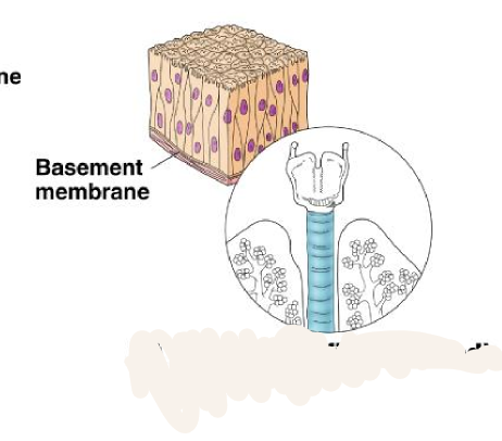
pseudostratified columnar epithelium
tissue that consists of a single layer of irregularly shaped and sized cells that give the appearance of multiple layers; found in ducts of certain glands and the upper respiratory tract
8
New cards
straified squamous
thick membrane
protects underlying tissues in areas subjected to abrasion
protects underlying tissues in areas subjected to abrasion
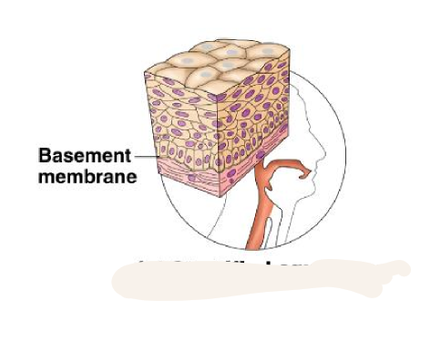
9
New cards
Transitions striated
shapes of cells that sketch lines organ of the urinary system
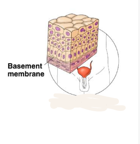
10
New cards
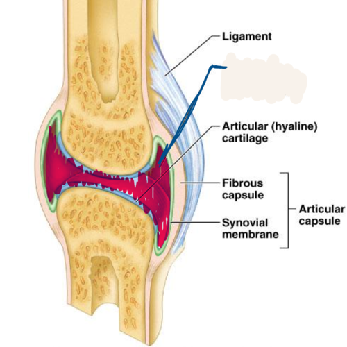
articular capsule
sleevelike structure around a synovical joint composed of a fibrous capsule and synovial membrane
this is called
this is called
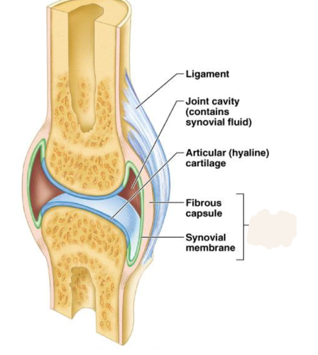
11
New cards
synovial membrane
membrane lining the capsule of a joint
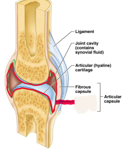
12
New cards
joint cavity
contains synovial fluid is called
13
New cards
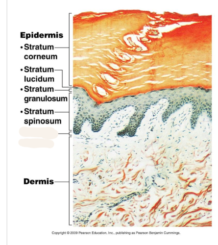
stratum basale
deepest layer of epidermis
14
New cards
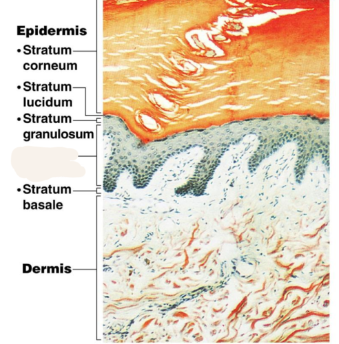
stratum spinosum
a layer of the epidermis that provides strength and flexibility to the skin
is called
is called
15
New cards
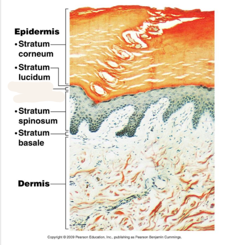
stratum granulosum (granular layer)
Layer of epidermis composed of cells that look like granules and are filled with keratin; replaces cells shed from stratum corneum
16
New cards
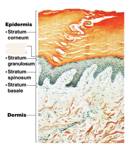
Stratum lucidum (clear layer)
Only in thick skin
Thin, translucent band superficial to the stratum granulosum
A few rows of flat, dead keratinocytes
Thin, translucent band superficial to the stratum granulosum
A few rows of flat, dead keratinocytes
17
New cards
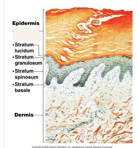
stratum corneum
outermost layer of epidermis
18
New cards
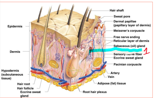
arrector pili muscle
An involuntary muscle fiber attached to the underside & base of the hair follicle
19
New cards
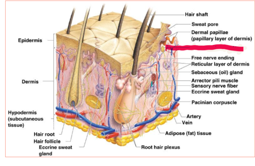
Meissner's corpuscles
sensitive touch receptors in the dermis
20
New cards
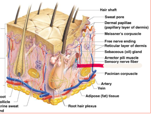
eccrine sweat glands
found in palms, soles of the feet, and forehead
21
New cards
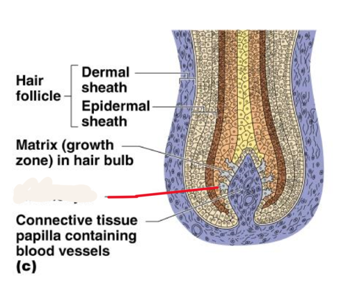
Melanocytes
cells that produce melanin
22
New cards
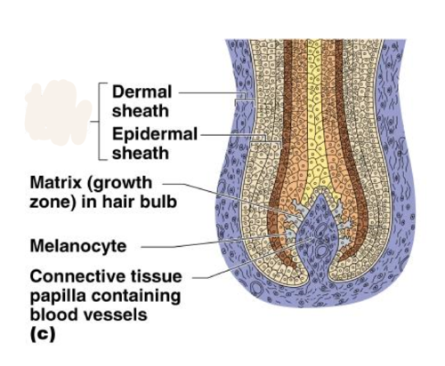
hair follicle
sac within which each hair grows
23
New cards
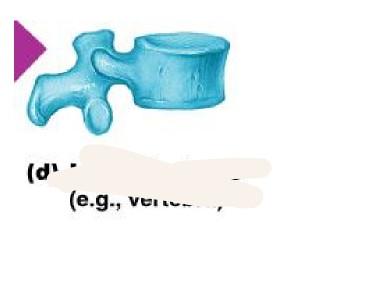
irregular bones
bones of the vertebrae and face
24
New cards
flat bones
bones of the ribs, shoulder blades, pelvis, and skull
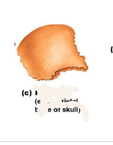
25
New cards
Central (Haversian) Canal
opening in the center of an osteon, carries blood vessels and nerves
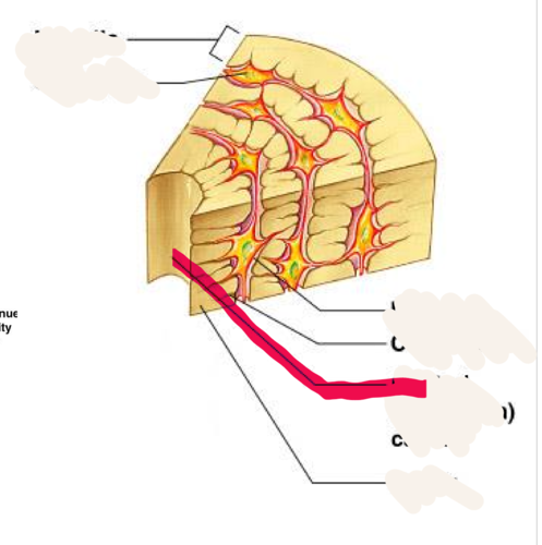
26
New cards
Canaliculus/canaliculi
tiny canals radiating outward from central canal
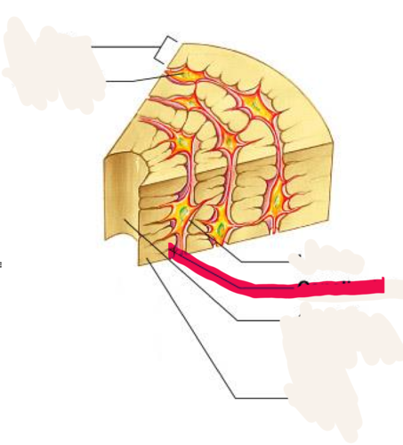
27
New cards
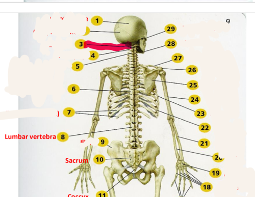
Axis (C2)
second cervical vertebrae
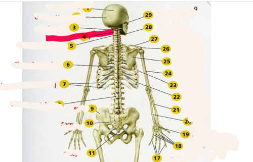
28
New cards
Atlas (C1)
supports the head
29
New cards
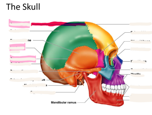
coronal suture
the suture between the parietal and frontal bones of the skull
30
New cards
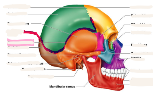
lambdoid suture
between parietal bones and occipital bone
31
New cards
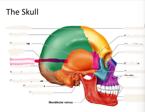
squamous suture
Between parietal and temporal bones
32
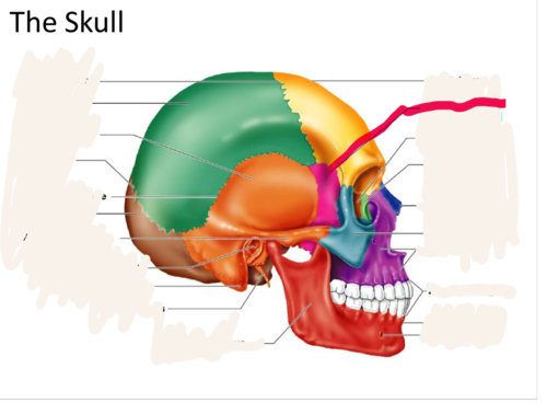
New cards
sphenoid bone
forms part of the base of the skull and parts of the floor and sides of the orbit
33
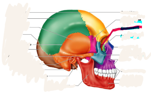
New cards
ethmoid bone
forms part of the posterior portion of the nose, the orbit, and the floor of the cranium
34
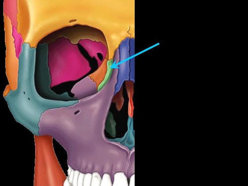
New cards
lacrimal bone
small fragile bone making up part of the front inner walls of each eye socket and providing room for the passage of the lacrimal ducts
35
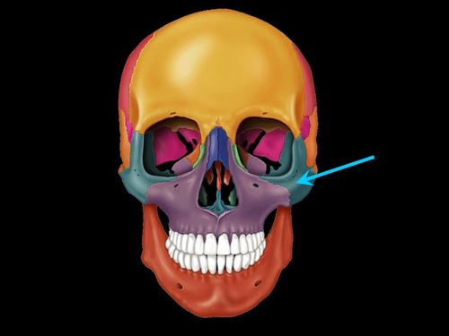
New cards
Zygomatic bone
cheek bone
36
New cards
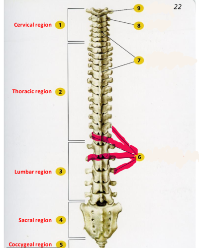
Transverse process of vertebra
forms joint with ribs in thoracic region, site for muscle attachment in lumbar region
37
New cards
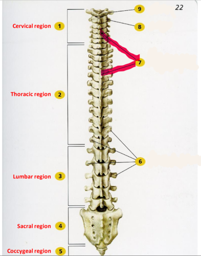
spinous process
sharp, slender projection
38
New cards
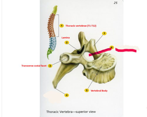
vertebral foramen
canal through which spinal cord passes
39
New cards
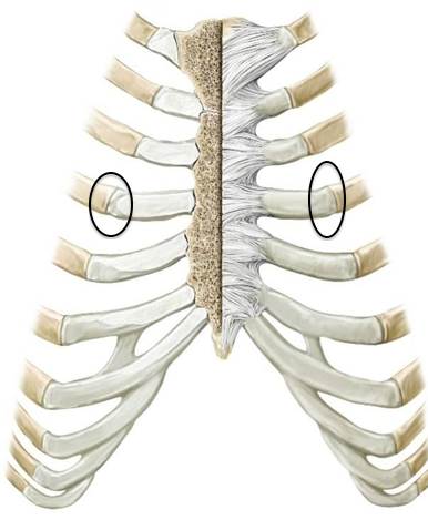
costochondral joint
where the cartilage meets the rib
40
New cards
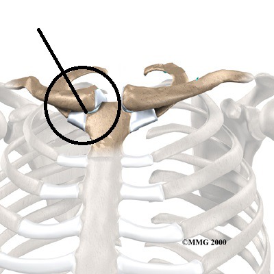
sternoclavicular joint
Articulation between the clavicle and the sternum
41
New cards
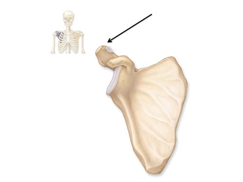
Acromion
Outward extension of the shoulder blade forming the point of the shoulder.
42
New cards
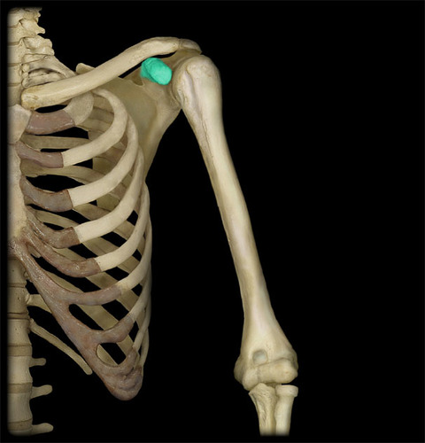
coracoid process
process above the glenoid cavity that permits muscle attachment
43
New cards
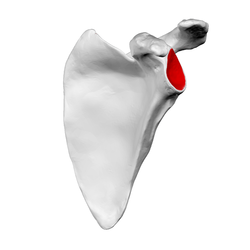
glenoid cavity
socket in scapular that receives head of humerus
44
New cards
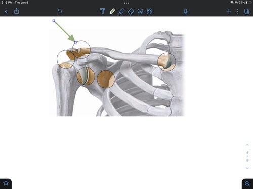
acrominoclavicular joint
the area where the clavicle and scapula connect
45
New cards
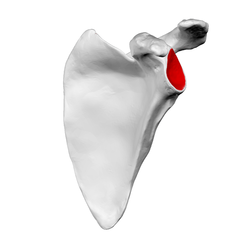
glenoid fossa of scapula
articulates with the head of the humerus
46
New cards
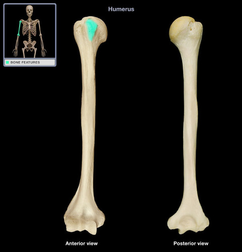
lesser tubercle
insertion of subscapularis muscle
47
New cards
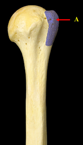
greater tubercle
Large lateral prominence; site of the attachment of rotator cuff muscles
48
New cards
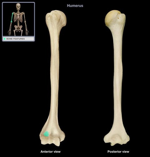
radial fossa of humerus
anterior depression that receives the radial head with flexed forearm
49
New cards
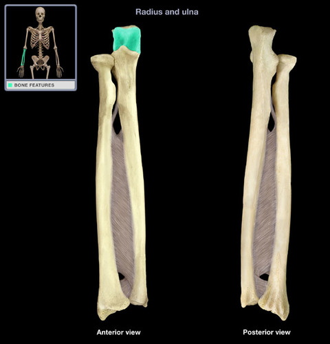
trochlear notch
articulates with trochlea of humerus
50
New cards
proximal radioulnar joint
head of radius articulates with radial notch of ulna

51
New cards
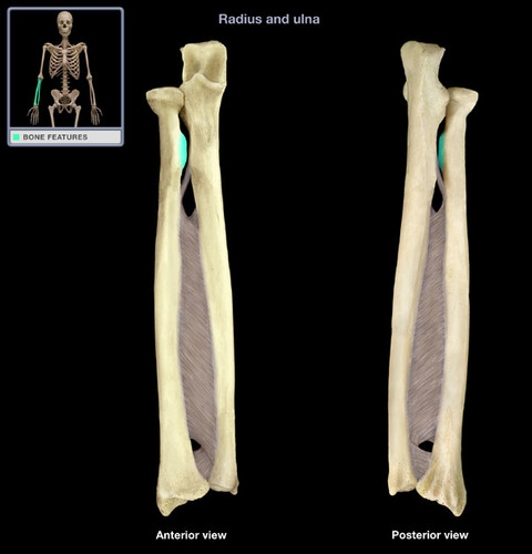
radial tuberosity
Name this specific part of the radius.
52
New cards
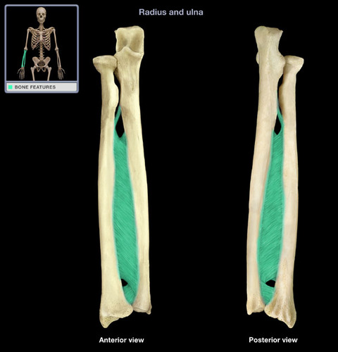
interosseous membrane
connects the tibia and fibula
53
New cards
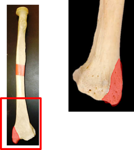
syloid process of radius
insertion of brachioradialis
54
New cards
distal radioulnar joint
head of ulna articulates with ulnar notch of radius

55
New cards
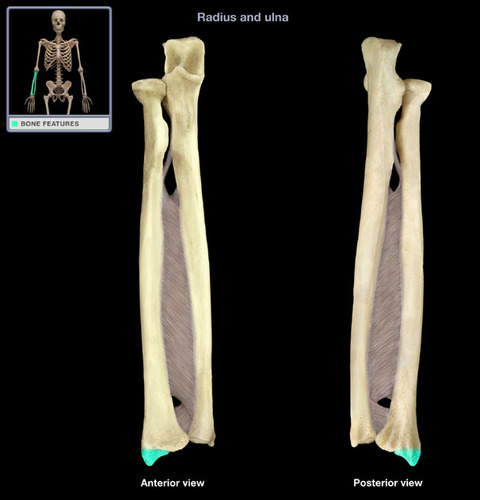
styloid process of ulna
Name the structure.
56
New cards
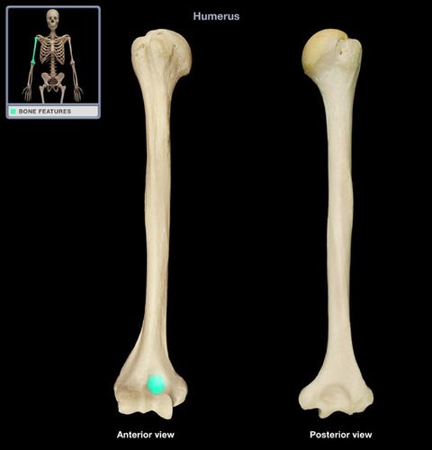
coronoid fossa
anterior depression that receives the coronoid process of the ulna during forearm flexion
57
New cards
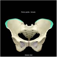
illiac crest
the upper curved edge of the ilium; has anterior (anterior iliac spine) and posterior projections
58
New cards
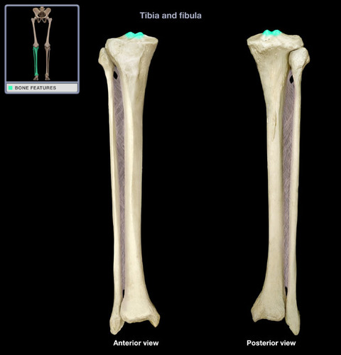
intercondylar eminence
irregular projection located between the two condyles
59
New cards
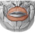
closes and protrudes lips is called the
orbicularis oris
60
New cards
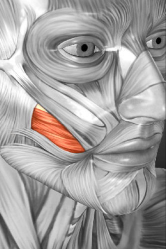
compresses cheek ( as in sucking), holds food between teeth during chewing. THis is called
buccinator
61
New cards
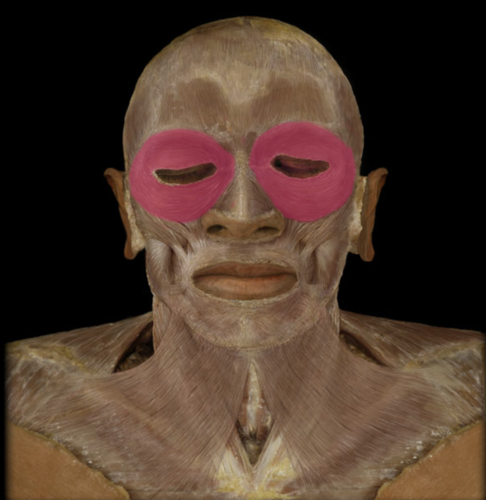
blinks and closes eyes, squints and winks
orbicularis oculi
62
New cards
extends from the corner of the mouth to the cheekbone
zygomaticus
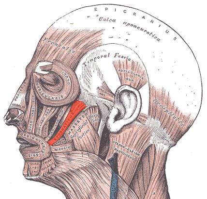
63
New cards
PULLS THE CORNERS OF THE MOUTH INFERIORLY
PRODUCING A DONWARD SAF OF THE MOUTH
PRODUCING A DONWARD SAF OF THE MOUTH
temporalis
64
New cards
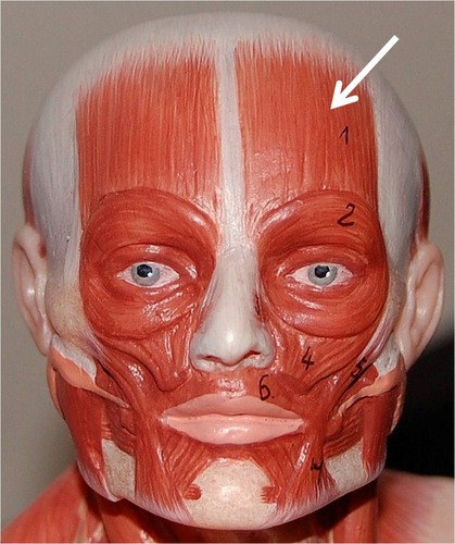
Frontalis
muscle of the forehead that moves the forehead skin and eyebrows
65
New cards
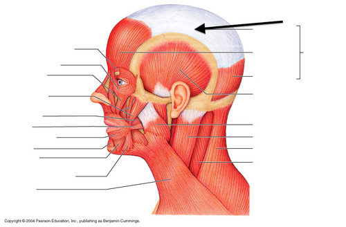
cranial aponeurosis
connects frontalis and occipitalis
66
New cards
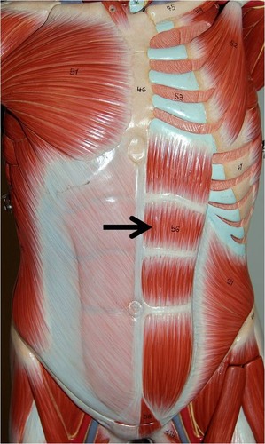
rectus abdominis
this type of muscle is called the
67
New cards
transversus abdominis
compresses abdomen, this is called the
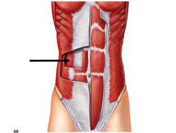
68
New cards
internal oblique
tenses abdominal wall and compresses abdominal contents. this is called
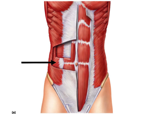
69
New cards
External onlique
flex and rotate vertebral column, this is called
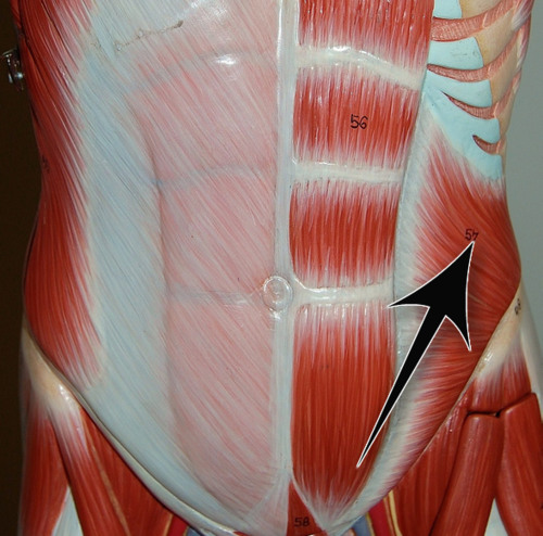
70
New cards
Brachialis
flexes forearm at elbow, this is called the
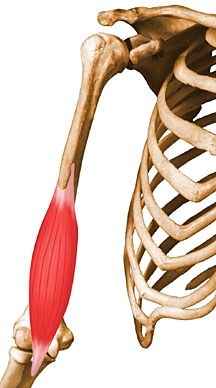
71
New cards
Brachioradialis
flexes forearm
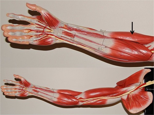
72
New cards
biceps brachii
Flexes and supinates forearm
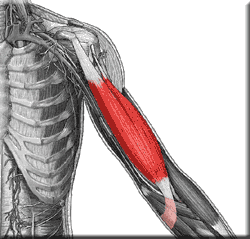
73
New cards
gluteus maximus
extends thigh
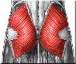
74
New cards
gluteus medius
abducts and medially rotates thigh
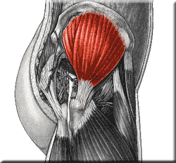
75
New cards
felxor carpi radialis
flexes and abducts wrist
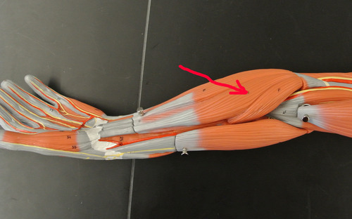
76
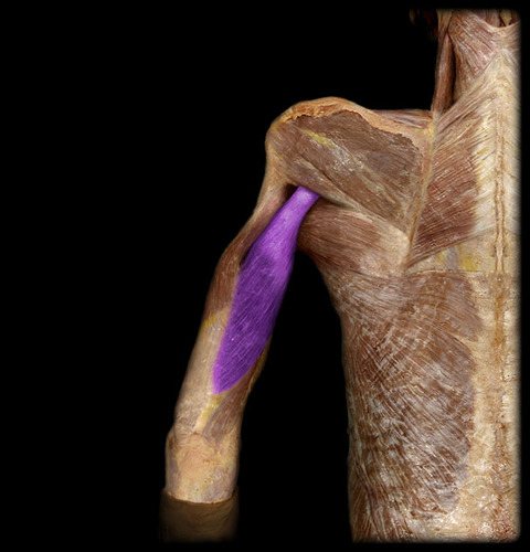
New cards
trieps brachii
insertions: olecranon of ulna
origin: shoulder girdle and proximal numerus
functions: extend elbow
origin: shoulder girdle and proximal numerus
functions: extend elbow
77
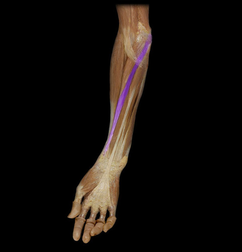
New cards
felxor carpi radialis
Origins: distal humerus
insertions: second and third metacarpals
functions: flexes wrist and abducts hands
insertions: second and third metacarpals
functions: flexes wrist and abducts hands
78
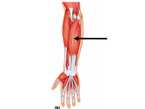
New cards
flexor digitorum superficialis
Orgin: distal humerus, ulna and radius
insertions: middle phalanges of second fifth finger
functions: flexes wrist and finger
insertions: middle phalanges of second fifth finger
functions: flexes wrist and finger
79
New cards
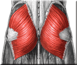
gluteus maximus
Functions: extends hip, abducts thigh; steadies pelvis during walking and flex knee and extend hip
insertions: proximal femur
origin: sacrum and ilium
insertions: proximal femur
origin: sacrum and ilium
80
New cards
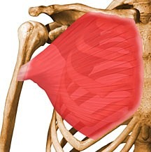
pectoralis major
Adducts and flexes humerus
81
New cards
unipennate muscle
all the muscle fibers are on the same side of the tendon

82
New cards
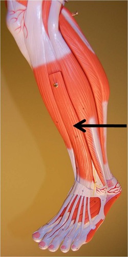
extensor digitorum longus
extends toes and dorsiflexes foot
83
New cards
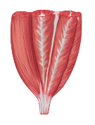
multipennate muscle
forms an angle with a tendon; do not move as far as parallel; contains more myofibrils than parallel muscles; develop more tension than parallel muscles; tendon branches within the muscle; ex: deltoid
84
New cards
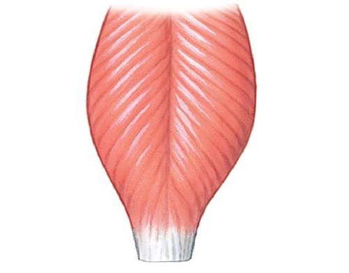
Bipennate
rectus femoris
85
New cards
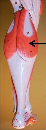
Gastrocnemius
Calf muscle
86
New cards
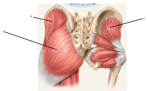
gluteal group muscles
gluteus maximus, gluteus medius, gluteus minimus
87
New cards
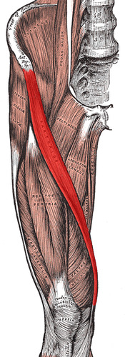
Sartorius
Flexes, abducts, and laterally rotates thigh at the hip; flexes knee
88
New cards
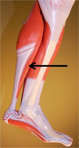
Soleus
plantar flexes foot
89
New cards
Illiopsoas
flexes hip

90
New cards
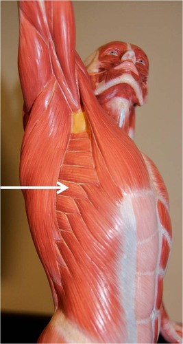
serratus anterior
pulls scapula anteriorly and downward
91
New cards
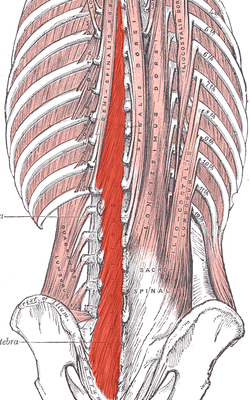
multifidus muscle
stabilizes lower thoracic and lumbar region of vertebral column
92
New cards
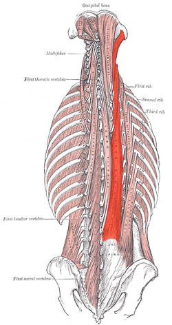
longissimus capitis
extends and rotates head
93
New cards
semispinalis capitis
extends head, bends head to one side, or rotates head
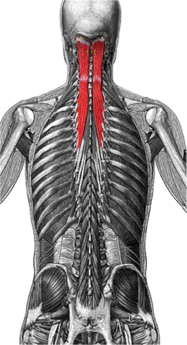
94
New cards
Flexion
bending a joint
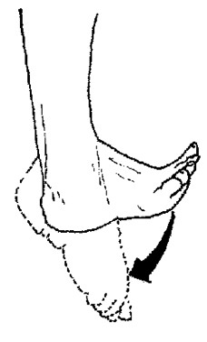
95
New cards
extension
Straightening of a joint
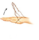
96
New cards
Rotation
CIRCULAR MOVEMENT AROUND AN AXIS
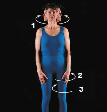
97
New cards
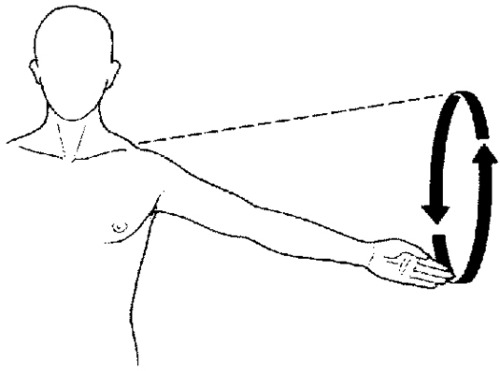
circumduction movement
moving the joint in a circular manner
98
New cards
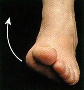
Inversion
turning inward
99
New cards
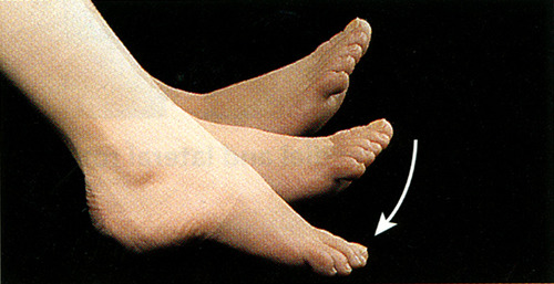
plantar flexion
bending of the sole of the foot by curling the toes toward the ground
100
New cards
Dosiflexion
bending of the foot or the toes upward
