Unit 2 : Integumentary System, Skeletal System, Histology
0.0(0)
Studied by 10 peopleCard Sorting
1/114
Last updated 1:31 AM on 10/5/22
Name | Mastery | Learn | Test | Matching | Spaced | Call with Kai |
|---|
No analytics yet
Send a link to your students to track their progress
115 Terms
1
New cards
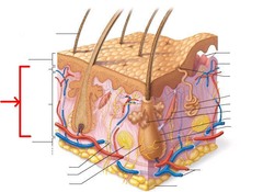
Integumentary System
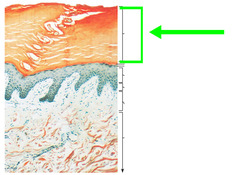
Covering of the body, consists of skin, hair, nails and sweat glands (sudoriferous) and oil glands (sebaceous).
2
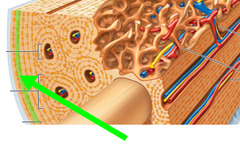
New cards
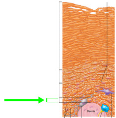
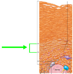
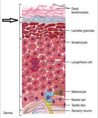
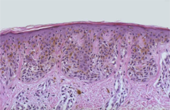
Epidermis
Superficial region. Sheet-like layers. Has epithelial stratified squamous cells and is dense connective. Nutrients from dermis diffuse into epidermis, giving them food.
3
New cards
Dermis
Underlines epidermis. Made up of loose & connective and dense connective tissue. Mostly fibrous connective tissue, vascular (rich in blood).
4
New cards
Hypodermis
Not technically apart of the skin because it is underneath the epidermis. It is the subcutaneous layer deep to the skin. Mostly made up of adipose tissue that absorbs shock and insulates. Anchors fat to underlying structures, mostly muscles.
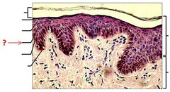
5
New cards
What does fat do?
Fat is fuel preserve and also insulates the body.
6
New cards
Keratinocytes
Most abundant epidermal cells. They produce water proofing keratin protein that gives skin its protective properties.
7
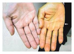
New cards
What are keratinocytes connected by and why?
Keratinocytes are connected by desmosomes which provide tensile strength.
8
New cards
Melanocytes
Deepest part of the epidermis. They produce melanin, which is packaged melanosomes. The melanosomes get transferred to the keratinocytes and protect nucleus from UV damage.
9
New cards
Why do the nuclei of mitotically active cells need melanosome protection?
Nuclei house DNA = mitosis. Harmful UV rays can cause unchecked cell growth and can cause cancer.
10
New cards
Dendritic (Langerhans Cells)
Star-shaped macrophages that patrol deepest epidermis.
11
New cards
What do dendritic cells do?
- Macrophages that eat and ingest invaders. They prevent entry of bacteria into the vasculatured dermis.
12
New cards
Tactile (Merkel) cells
Sensory touch receptors that sense touch. They can feel light or fine touh micromovements.
13
New cards
Difference between thick and thin skin?
Thick skin has 5 layers (which include stratum lucidum) and thin skin only has 4 layers.
14
New cards
What are the five layers of skin? From most deep layer to most superficial.
Stratum basale, Stratum Spinosum, Stratum granulosum, Stratum lucidum, Stratum corneum.
15
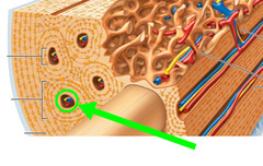
New cards
Stratum basale (basal layer)
-Deepest epidermal layer firmly attached to the dermis
single row of stems. The layer right before the hypodermis.
-The single row of stem cells are highly mitotic and actively producing and dividing.
-10-25% of this layer is made up of melanocytes.
single row of stems. The layer right before the hypodermis.
-The single row of stem cells are highly mitotic and actively producing and dividing.
-10-25% of this layer is made up of melanocytes.
16
New cards
Why do the stem layers of stratum basale never deplete?
As the cells are mitotically active, when they divide, one daughter cell remains while the other migrates to the apical surface. It takes about 25-45 days.
17
New cards
Stratum spinosum (prickly/spiny layer)
- Several layers thick (where cells start to become dehydrated.
- Cell contain web-like system of intermediate prekeratin filaments attached to desmosomes.
- Desmosomes allow cells to resist tension and pulling.
- Keratinocytes in this layer appear prickly/spikey.
- Cell contain web-like system of intermediate prekeratin filaments attached to desmosomes.
- Desmosomes allow cells to resist tension and pulling.
- Keratinocytes in this layer appear prickly/spikey.
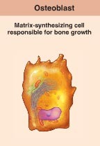
18
New cards
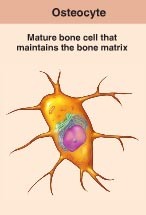
Stratum granulosum (granular layer)
Four to six cells thick, but the cells are flattened so layer = thin cell keratinization begins.
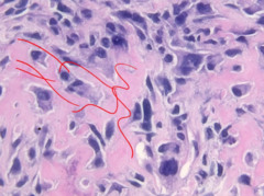
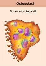
- Cell nuclei/organelles begin to disintegrate.
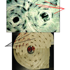
- Cells accumulate keratinohyaline that form keratin fibers.
- Accumulate lamellar granules.
- Any cells above this layer die because they are too far from dermal capillaries.
- Cell nuclei/organelles begin to disintegrate.
- Cells accumulate keratinohyaline that form keratin fibers.
- Accumulate lamellar granules.
- Any cells above this layer die because they are too far from dermal capillaries.
19
New cards
Stratum lucidum
Only found in thick skin. The clear because there are no nuclei. The cells are now dead.
- 2/3 flat rows.
- 2/3 flat rows.
20
New cards
Stratum corneum
Absent of nuclei, anucleated, keratinized dead layer of cells.
- 20/30 flat rows
- physical barrier to protect deeper cells from environment, prevent water loss, protects against abrasion and penetration.
- Acts as a barrier against biological, chemical and physical assaults.
- 20/30 flat rows
- physical barrier to protect deeper cells from environment, prevent water loss, protects against abrasion and penetration.
- Acts as a barrier against biological, chemical and physical assaults.
21
New cards
What is apoptosis?
Programmed cell death. Dead cells in the skin slough off as dandruff and dead skin.
22
New cards
What is the Dermis?
- strong flexible connective tissue beneath the epidermis. It is made up of fibers in matrix that bind body together.
- Contains nerves, blood vessels and lymphatic vessels (vascularized).
- Contains nerves, blood vessels and lymphatic vessels (vascularized).
23
New cards
What kind of cells are within the dermis?
Fibroblasts, macrophages and occasionally mast cells and white blood cells.
24
New cards
What do fibroblasts do?
Secrete cell matrix, collagen fibers and active stem cells.
25
New cards
What are macrophages and what do they do?
>Macrophages are large phagocytic cells that develop from some leucocytes
>Some wander through tissues looking for pathogens and destroying them
>Others have a fixed place and destroy pathogens that come to them
- Macrophages phagocytize and pinocytize.
>Some wander through tissues looking for pathogens and destroying them
>Others have a fixed place and destroy pathogens that come to them
- Macrophages phagocytize and pinocytize.
26
New cards
What is the papillary layer?
The thinner layer of loose and airy areolar connective tissue of interlacing collagen, elastic fibers and blood vessels.
27
New cards
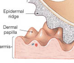
What are dermal papillae?
Superficial region of dermis that sense fingerlike projections up into epidermis.
- in thick skin, it lies on top of dermal ridges, which gives rise to friction ridges (finger prints!)
- in thick skin, it lies on top of dermal ridges, which gives rise to friction ridges (finger prints!)
28
New cards
What is the friction ridge for?
Enhances gripping ability and contributes to the sense of touch. Sweat pores in ridges leave unique fingerprint-like pattern.
29
New cards
What do projections in the dermal papillae contain?
- Capillary loops, free nerve endings and touch receptors.
30
New cards
Does location of capillary loops, free nerve endings and touch receptors matter?
Yes! If found deeper in dermis, it detects deeper pressure, but if it is superficial, it will detect fine touch.
31
New cards
What is the reticular layer?
-makes up ~80% of dermal thickness.
- Consists of coarse, dense, fibrous connective tissue.
- Elastic fibers = stretch and recoil properties while collagen = tensile strength.
- Layer contains spillage of adipose tissue.
- Consists of coarse, dense, fibrous connective tissue.
- Elastic fibers = stretch and recoil properties while collagen = tensile strength.
- Layer contains spillage of adipose tissue.

32
New cards
How do dermal modifications result in characteristic markings?
Surgeons are able to use specific lines on the body cause by collagen fibers in order to cut along and prevent scarring.
33
New cards
Cleavage (tension lines)
Caused by collagen fibers running parallel to skin surface.
34
New cards
Flexure line
Adheres skin/dermis to joints where it is adhered to deeper structures.
35
New cards
What causes skin color?
- Melanin, Carotene & Hemoglobin
36
New cards
Melanin
- Brownish color made in skin by melanocytes within the stratum basale.
- The more sun you are exposed to, the more melanin your skin secretes.
- Packages melanosomes to the keratinocytes to shield the nucleus from UV sunlight.
- The more sun you are exposed to, the more melanin your skin secretes.
- Packages melanosomes to the keratinocytes to shield the nucleus from UV sunlight.
37
New cards
Carotene
- Yellow-orange pigment
- Most obvious in palms and soles.
Accumulates to stratum corneum + hypodermis.
- Can be converted to Vitamin A for vision and epidermal health.
- Most obvious in palms and soles.
Accumulates to stratum corneum + hypodermis.
- Can be converted to Vitamin A for vision and epidermal health.
38
New cards
Hemoglobin
- Reddish pigment found in our RBCs.
- In lighter complexioned individuals, pinkish hue of fair skin is due to lower levels of melanin.
- In lighter complexioned individuals, pinkish hue of fair skin is due to lower levels of melanin.
39
New cards
Why do caucasian individuals have transparent skin?
- The amount of melanocytes that people have in the skin are the same, however, the amount of melanin an individual secretes is entirely different.
- Caucasian individuals are more likely to be transparent and hemoglobin shows through more.
- Caucasian individuals are more likely to be transparent and hemoglobin shows through more.
40
New cards
Eccrine (merocrine) sweat gland
- released to the exterior and is everywhere.
- abundant on palms, soles and forehead.
- function is thermoregulation
- Secretes sweat and is devoid of odor
- abundant on palms, soles and forehead.
- function is thermoregulation
- Secretes sweat and is devoid of odor
41
New cards
Apocrine sweat gland
- Stinky sweat glands that are most confined to axillary (armpit)and urogenital areas (areas with pubic hair)
- Sweat secretion is viscous milky or yellowish sweat that contains fatty substances and proteins.
- Begins to secrete at puberty.
- Sweat secretion is viscous milky or yellowish sweat that contains fatty substances and proteins.
- Begins to secrete at puberty.
42
New cards
Why is apocrine sweat smelly?
Bacteria break down fatty substance/viscous protein sweat, leading to body odor because ammonia is release.
- Remember that protein is made up of amino acids that have the N group.
- Remember that protein is made up of amino acids that have the N group.
43
New cards
Sebaceous (oil) glands
Body relies on sebum to lubricate skin and hair follicles.
- Androgens activate sebaceous glands to be active @ PUBERTY.
- Sebaceous glands self destruct after a cell secretes.
- Androgens activate sebaceous glands to be active @ PUBERTY.
- Sebaceous glands self destruct after a cell secretes.
44
New cards
Arrector pili muscle
An involuntary muscle fiber attached to the underside & base of the hair follicle.
- Causes the hair on your epidermis to stand up
- Causes the hair on your epidermis to stand up
45
New cards
What are some function of skin?
- chemical, physical and biological barrier.
- Skin protects against microorganisms, abrasions, temperature extremes and harmful chemicals.
- Skin protects against microorganisms, abrasions, temperature extremes and harmful chemicals.
46
New cards
Skin : Chemical barrier
-Secretes sweat (antimicrobial proteins), sebum & defensins that kill bacteria.
- Low pH of skin retards bacterial manipulation because bacteria cell wall is negative.
- Low pH of skin retards bacterial manipulation because bacteria cell wall is negative.
47
New cards
Skin : Physical barrier
- Glycolipids within the stratum corneum block most water and water-soluble lipids.
- Skin takes in lipid soluble substances like hydrocortisone because it is made of steroids = lipid soluble.
- Skin takes in lipid soluble substances like hydrocortisone because it is made of steroids = lipid soluble.
48
New cards
Skin : Biological barrier
- Epidermis contains phagocytic cells.
- Dendritic cells engulf antigens and present them to WBC to launch and activate immune response system.
- Dermis contains macrophages (phagocytic WBC)
- Dendritic cells engulf antigens and present them to WBC to launch and activate immune response system.
- Dermis contains macrophages (phagocytic WBC)
49
New cards
Body Temperature Regulation : Insensible respiration
Resting body temperature, you sweat about 500 ml a day of unnoticeable sweat.
50
New cards
Body Temperature Regulation : Sensible respiration
Body temperature rises because of the dilation of dermal vessels and can increase sweat glands activity to produce 12 L of sweat.
51
New cards
Blood vessel dilation
- When blood vessels are brought closer to the dermis of the skin and the body gets hotter. Occurs when you're feeling hot so you can sweat (sweat cools the body).
52
New cards
Blood vessel constriction
- When blood vessels move further into the skin so heat cannot escape. When you're feeling cold.
53
New cards
Cutaneous sensory receptors
Part of nervous system. Exteroreceptors respond to stimuli outside body such as temperature and touch.
Free nerve endings sense painful stimuli.
Free nerve endings sense painful stimuli.
54
New cards
Metabolic function of skin
- Synthesis of vitamin D needed for calcium absorption in intestine.
- Keratinocytes can activate some hormones (convert cortisone into hydrocortisone).
- Skin makes collagenase to aid in turnover of wrinkles
- Keratinocytes can activate some hormones (convert cortisone into hydrocortisone).
- Skin makes collagenase to aid in turnover of wrinkles
55
New cards
Skin / Reservoir of Blood
- Skin holds 5% of body's blood volume.
- Skin vessels can be restricted to shunt (redirect) blood to other organs.
- Skin vessels can be restricted to shunt (redirect) blood to other organs.
56
New cards
Skin / Excretion
- Skin can secrete limited amounts of waste such as ammonia, urea and uric acid.
-Sweat = salt and water loss
-Sweat = salt and water loss
57
New cards
Skeletal cartilage
made of highly resilient, molded cartilage tissue that consists primarily of water.
Contains no blood vessels or nerves
Contains no blood vessels or nerves
58
New cards
Perichondrium
Layer of dense connective tissue surrounding cartilage.
- Helps cartilage resist outward expansion.
- Contains blood vessels to diffuse nutrient delivery to the cartilage.
- Strips of dense connective tissue.
- Helps cartilage resist outward expansion.
- Contains blood vessels to diffuse nutrient delivery to the cartilage.
- Strips of dense connective tissue.
59
New cards
Chondrocytes
Cells encased in small cavities (lacunae). They secrete the matrix of cartilage and become embedded into it.
60
New cards
Hyaline Cartilage
Provides support, flexibility & resilience.
- Most abundant, collagen only.
- Found in articular joints, costal ribs, respiratory tract and nasal.
- Most abundant, collagen only.
- Found in articular joints, costal ribs, respiratory tract and nasal.
61
New cards
Elastic cartilage
Contains elastic fibers, support, flexibility and stability.
- Found in external ear and epiglottis.
- Found in external ear and epiglottis.
62
New cards
Fibrocartilage
Thick collagen fibers for great tensile strength.
- Found in areas with discs such as the knee and vertebral discs
- Found in areas with discs such as the knee and vertebral discs
63
New cards
Appositional growth
Perichondrium secretes matrix to widen the girth of bone.
64
New cards
Interstitial growth
Chondrocytes divide and secrete new matrix expanding the length of bone.
65
New cards
Calcification of cartilage
deposit of salt that hardens cartilage.
- Not yet bone, to become bone it must ossify.
- Not yet bone, to become bone it must ossify.
66
New cards
Axial Skeleton
Long axis of body, skull, vertebral column and rib cage
67
New cards
Appendicular Skeleton
Bones of upper and lower limbs, girdles and skeletal girdle.
- Hip & clavicle + scapula.
- Hip & clavicle + scapula.
68
New cards
Long bone
Longer than they are wide.
- Limb bones and phalanges.
- Limb bones and phalanges.
69
New cards
Short bone
Cube shaped, width and length are equal.
- Found in wrist and ankles
- Found in wrist and ankles
70
New cards
Sesamoid
Small seed like shape, form within tendons (only example is patella)
71
New cards
Flat bone
thin, flat, slightly curved
- Found in sternum, scapulae, ribs and most skull bones
- Found in sternum, scapulae, ribs and most skull bones
72
New cards
Irregular bone
complicated shapes
- Found in vertebrae + hip bones, sacrum
- Found in vertebrae + hip bones, sacrum
73
New cards
Compact bone
Dense outer layer on every bone that is smooth or solid
74
New cards
Spongy bone
made up of a honey comb. of small needle-like or flat pieces of bone called trabeculae
75
New cards
Periosteum
Covers the outside of the compact bone except joints.
76
New cards
Fibrous layer
Outer layer consists of dense irregular connective tissue
77
New cards
Osteogenic layer
Inner layer of abutting bone that contains primitive osteogenic stem cells that gives rise to most all bones
78
New cards
Endosteum
membranous lining of the hollow cavity of the inside of the compact bone
79
New cards
Where is red bone marrow found?
Scattered throughout spongy bone (diploe). No defined marrow cavity.
80
New cards
Where is yellow bone marrow found?
It is found within the yellow bone marrow cavity.
81
New cards
Diaphysis
Tubular shaft that forms long axis of bone. Contains medullary cavity of yellow marrow
82
New cards
Epiphyses
Ends of long bones that consist of compact bone externally and spongy bone internally.
83
New cards
Epiphyseal line
Ossified plate between diaphysis and epiphysis.
- Remnants where bone growth begins to occur.
- Remnants where bone growth begins to occur.
84
New cards
If there is no epiphyseal line, what does that mean?
Growth plate means growth is still possible! Epiphyseal line may not be found in children who are still maturing.
85
New cards
Hematopoietic tissue in bones
As you age, your red bone marrow converts into yellow bone marrow.
86
New cards
What bone marrow do adults have?
Primarily yellow bone marrow.
87
New cards
What bone marrow do children have?
Primarily red bone marrow.
88
New cards
What happens if an adult / vperson if anemic?
Yellow bone marrow can convert to red.
89
New cards
Osteogenic cells
Mitotically active stem cells in periosteum and endosteum. When stimulated, they can differentiate into osteoblasts or bone-lining cells.
90
New cards
Osteoblasts
Bone-forming cells that secrete unmineralized bone matrix called osteoid. They are mitotically active.
91
New cards
Osteoid
Made up of collagen and calcium-binding proteins.
92
New cards
Osteocytes
Immature bone cells in lacunae that no longer divide. They maintain bone matrix and act as stress or strain sensors.
- Respond to mechanical stimuli such as increased force on bone or weightlessness.
-Communicate to osteoblasts and osteoclasts so bone remodeling can occur.
- Connected through gap junctions.
- Deposit actual calcium into the matrix.
- Respond to mechanical stimuli such as increased force on bone or weightlessness.
-Communicate to osteoblasts and osteoclasts so bone remodeling can occur.
- Connected through gap junctions.
- Deposit actual calcium into the matrix.
93
New cards
Bone-lining cells
On external bone called : periosteal cells.
On internal bone called : endosteal cells.
On internal bone called : endosteal cells.
94
New cards
Osteoclasts
Hemopoietic stem cells merge to become osteoclasts, they have numerous amounts of nuclei.
- Osteoblasts send them signals in order to stay alive, if not they perform cell death / apoptosis.
- When active, cells are located in depressions called reabsorption bays.
- Function in bone resorption.
- Cells have ruffled borders that serve to increase surface area.
- Addition of acid to ionize calcium phosphate in bone matrix.
95
New cards
Osteon
Structural unit of compact bone.
- Consists of elongated cylinder that runs parallel to long bone axis.
- Lamellae contains collagen fibers that run in different directions in adjacent rings in order to resist twisting and withstand stress.
- Consists of elongated cylinder that runs parallel to long bone axis.
- Lamellae contains collagen fibers that run in different directions in adjacent rings in order to resist twisting and withstand stress.
96
New cards
Canaliculi
Hairlike canals that connect lacunae to eachother and osteocytes via cell projections. Allows communication between all osteocytes of osteon and permits nutrients and wastes to be relayed.
97
New cards
Interstitial Lamellae
fill spaces between osteons
98
New cards
Circumferential lamellae
located deep to periosteum and superficial to endosteum and extend around entire circumference of the diaphysis and resist twisting of long bone
99
New cards
Haversian canal
one of a network of tubes running through compact bone that contains blood vessels and nerves
100
New cards
Organic components of bone
Osteogenic cells, osteoblasts, osteocytes, bone-lining cells, osteoclasts and osteoids.