AP Biology Unit 2: Comprehensive Study Notes
Cell Structure: Subcellular Components
To understand biology, one must understand the fundamental unit of life: the cell. The cell is not just a fluid-filled sack; it is a complex factory with specialized machinery. Understanding the structure-function relationship is the core theme of this unit.
Ribosomes: The Protein Factories
Ribosomes are the cellular distinct machinery responsible for protein synthesis. They are composed of ribosomal RNA (rRNA) and protein.
- Key Feature: Ribosomes are not membrane-bound. Because of this, they are found in all forms of life (Eukaryotes and Prokaryotes), reflecting the common ancestry of all known life.
- Free Ribosomes: Floating in the cytosol. They generally synthesize proteins that function within the cytosol.
- Bound Ribosomes: Attached to the Rough ER/Nuclear Envelope. They synthesize proteins usually destined for insertion into membranes, packaging within organelles (like lysosomes), or export (secretion) from the cell.
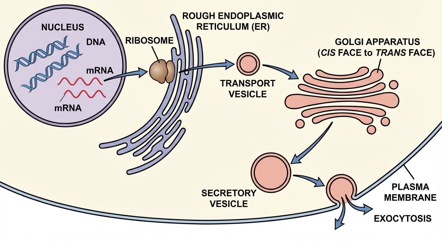
Endoplasmic Reticulum (ER)
The ER is a network of membranous sacs and tubes.
- Rough ER:
- Structure: Network of membrane tubes studded with ribosomes (hence "rough").
- Function: compartmentalizes the cell; facilitates the correct folding of newly synthesized proteins; adds carbohydrate chains to proteins (glycoproteins); keeps secretory proteins separate from cytosolic proteins.
- Smooth ER:
- Structure: Network of membrane tubes lacking ribosomes.
- Function: Synthesizes lipids (oils, steroids, phospholipids); metabolizes carbohydrates; detoxifies drugs and poisons (often liver cells are packed with Smooth ER); stores Calcium ions ($Ca^{2+}$) for muscle contraction.
The Golgi Complex
Think of the Golgi as the warehouse and shipping center (UPS/FedEx) of the cell.
- Structure: A series of flattened membranous sacs called cisternae.
- Function:
- Correct folding and chemical modification of newly synthesized proteins (adding molecular tags).
- Packaging proteins for trafficking to other parts of the cell or for exocytosis (leaving the cell).
- Directionality: The cis face receives vesicles from the ER; the trans face ships vesicles out.
Mitochondria
The powerhouse of the cell, responsible for cellular respiration.
- Structure: Double membrane. The outer membrane is smooth, but the inner membrane is highly convoluted, forming folds called cristae.
- Function: Synthesizes ATP via the Krebs cycle and electron transport chain.
- Significance of Folding: The cristae drastically increase the surface area of the inner membrane, allowing for more ATP synthase enzymes and electron transport chains, thereby maximizing energy production efficiency.
Chloroplasts
Found in specialized photosynthesizing organisms (plants, algae).
- Structure: Double outer membrane. Inside, there are membrane-bound sacs called thylakoids. Thylakoids are stacked into grana.
- Stroma: The fluid surrounding the thylakoids (contains chloroplast DNA and ribosomes).
- Function: Carbon fixation (Calvin-Benson cycle) occurs in the stroma; light-dependent reactions occur in the thylakoid membranes.
Lysosomes and Vacuoles
- Lysosomes: Membrane-enclosed sacs containing hydrolytic enzymes. They digest macromolecules, recycle the cell's own organic materials (autophagy), and facilitate programmed cell death (apoptosis).
- Vacuoles: Large membrane-bound vesicles. In plants, the large central vacuole helps maintain turgor pressure (keeping the plant upright) and stores water, ions, and waste.
Cell Size
Cells are generally small. This is not a coincidence; it is a mathematical necessity dictated by the Surface Area-to-Volume Ratio ($SA:V$).
The Mathematical Limitation
For a cell to survive, it must exchange materials (nutrients in, waste out) with its environment across the cell membrane.
- Surface Area ($SA$): The amount of membrane available for exchange.
- Volume ($V$): The internal amount of cytoplasm requiring nutrients and producing waste.
As a cell increases in size, its volume grows proportionally faster than its surface area.
As side length ($s$) increases, the ratio $s^2/s^3$ decreases clearly.
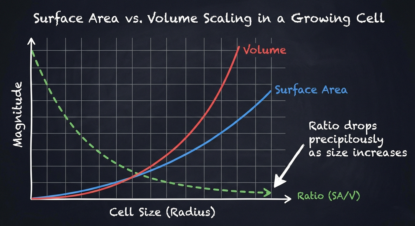
Implications for Function
- High $SA:V$ Ratio: Desirable. Allows efficient diffusion of materials. Small cells have high ratios.
- Low $SA:V$ Ratio: Undesirable. If a cell gets too big, the center cannot receive nutrients fast enough, and waste builds up to toxic levels.
Adaptations to Increase Ratio
Complex organisms don't just have bigger cells; they have more cells. However, specialized cells often modify their shape to increase Surface Area without significantly increasing Volume:
- Root Hairs: In plants, maximize water absorption.
- Microvilli: In the small intestine, tiny projections on epithelial cells increase absorption of nutrients.
- Flattened Shapes: Red blood cells or flatworms.
Cell Compartmentalization
Compartmentalization refers to the presence of membrane-bound organelles that create separate environments within the cell.
Why Compartmentalize?
- Metabolic Efficiency: Enzymes and substrates for specific processes remain concentrated in one area (e.g., Krebs cycle enzymes inside the mitochondrial matrix).
- Incompatible Reactions: Separating opposing reactions. For example, keeping hydrolytic enzymes (which digest proteins) inside the lysosome prevents them from digesting the cell's own cytosol.
- Specialized Environments: Membranes allow organelles to maintain specific internal conditions. For instance, lysosomes maintain a highly acidic pH (~5) which differs from the neutral cytosol (~7.2).
Prokaryotes vs. Eukaryotes
- Prokaryotes (Bacteria/Archaea): Generally lack internal membrane-bound organelles. However, they still have internal regions with specialized structures and functions (e.g., nucleoid region).
- Eukaryotes (Animals/Plants/Fungi/Protists): Highly compartmentalized via the endomembrane system.
Origins of Cell Compartmentalization
How did eukaryotic cells evolve from simpler ancestors? The leading explanation is the Endosymbiotic Theory.
The Endosymbiotic Theory
This theory proposes that membrane-bound organelles (specifically mitochondria and chloroplasts) evolved from free-living prokaryotic cells that were engulfed by an ancestral anaerobic eukaryote.
- The Mitochondrion Story: An ancestral cell engulfed an oxygen-using, non-photosynthetic prokaryote. Instead of digesting it, the host formed a symbiotic relationship. The host provided protection/nutrients; the guest provided ATP. This "guest" became the mitochondrion.
- The Chloroplast Story: A cell (already containing mitochondria) engulfed a photosynthetic prokaryote (cyanobacteria), which became the chloroplast.
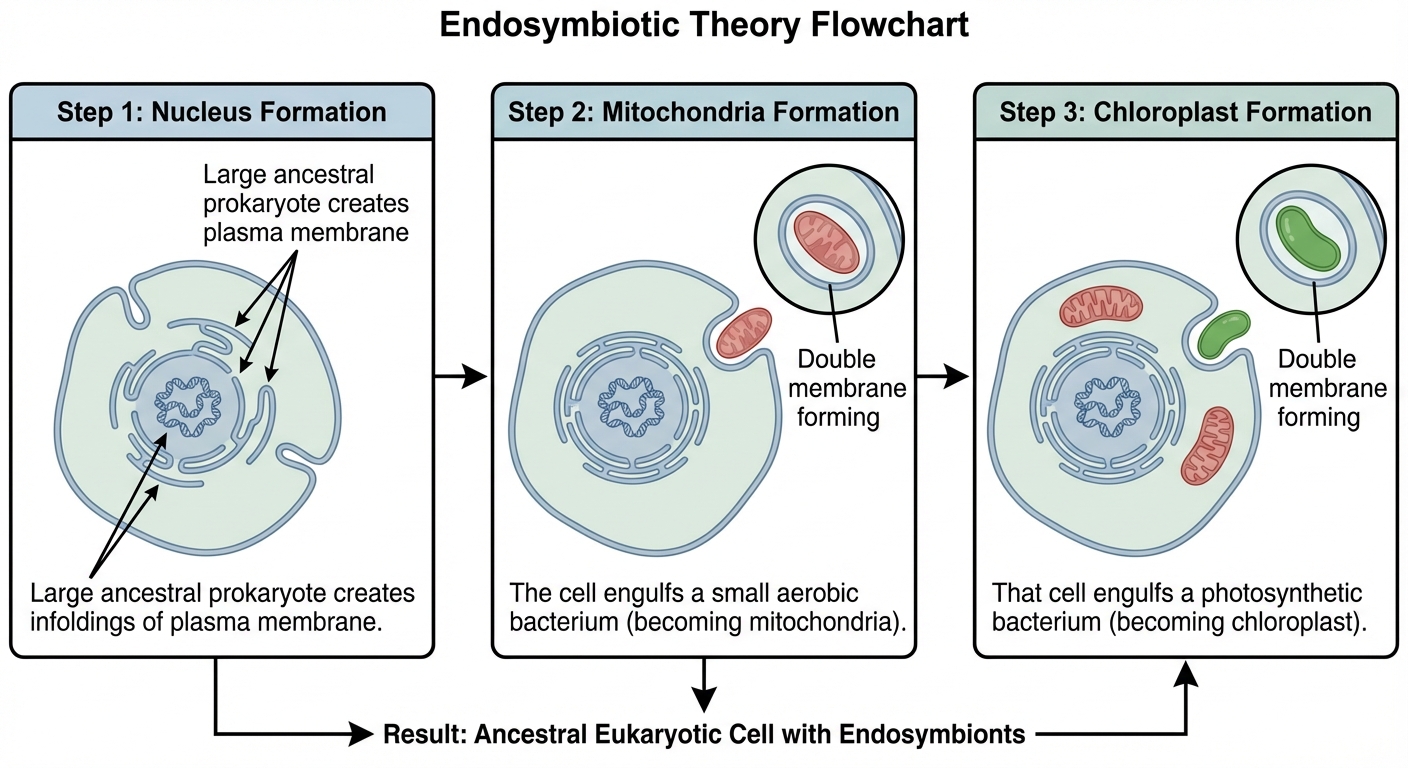
The Evidence (Mnemonic: DR. MD)
There is substantial structural evidence supporting this relationship between mitochondria/chloroplasts and bacteria:
- Division: Mitochondria and chloroplasts reproduce independently of the cell via binary fission (just like bacteria).
- Ribosomes: They contain their own ribosomes, which are 70S (bacterial size), not 80S (eukaryotic size).
- Membranes: They have double membranes. The inner membrane matches the composition of bacterial membranes; the outer membrane matches the eukaryotic host.
- DNA: They possess their own circular DNA, distinct from the linear DNA in the nucleus.
Common Mistakes & Pitfalls
- Confusing Cell Wall with Cell Membrane:
- Correction: All cells have a plasma membrane. Only plants, fungi, and some prokaryotes/protists have cell walls. The membrane controls active transport; the wall provides structural support.
- Misunderstanding "Bound" Ribosomes:
- Mistake: Thinking bound ribosomes are permanently stuck to the ER.
- Correction: Ribosomes cycle between free and bound states depending on the protein they are synthesising at the moment.
- Smooth ER Function:
- Mistake: Thinking Smooth ER makes proteins because it is "ER".
- Correction: Smooth ER makes lipids and detoxifies. Rough ER handles proteins.
- SA:V Calculation Interpretation:
- Mistake: Thinking a large cell has a large SA:V ratio.
- Correction: As size goes UP, the ratio goes DOWN. The most efficient cell is the small one.