AP Biology Unit 4: Mechanisms of Cell Signaling and Transduction
Overview of Cell Communication
Cell communication is the biological process by which cells detect and respond to signals in their environment. This is critical for maintaining homeostasis, coordinating growth, and allowing multicellular organisms to function as a unified entity. The fundamental unit of this communication is the interaction between a ligand (signaling molecule) and a receptor.
Evolutionary Significance
Signal transduction pathways are highly conserved across evolutionary history. The similarities in pathways between diverse organisms (e.g., yeast mating factors and mammalian growth factors) suggest that these mechanisms evolved in a common ancestor long ago.
Types of Signaling
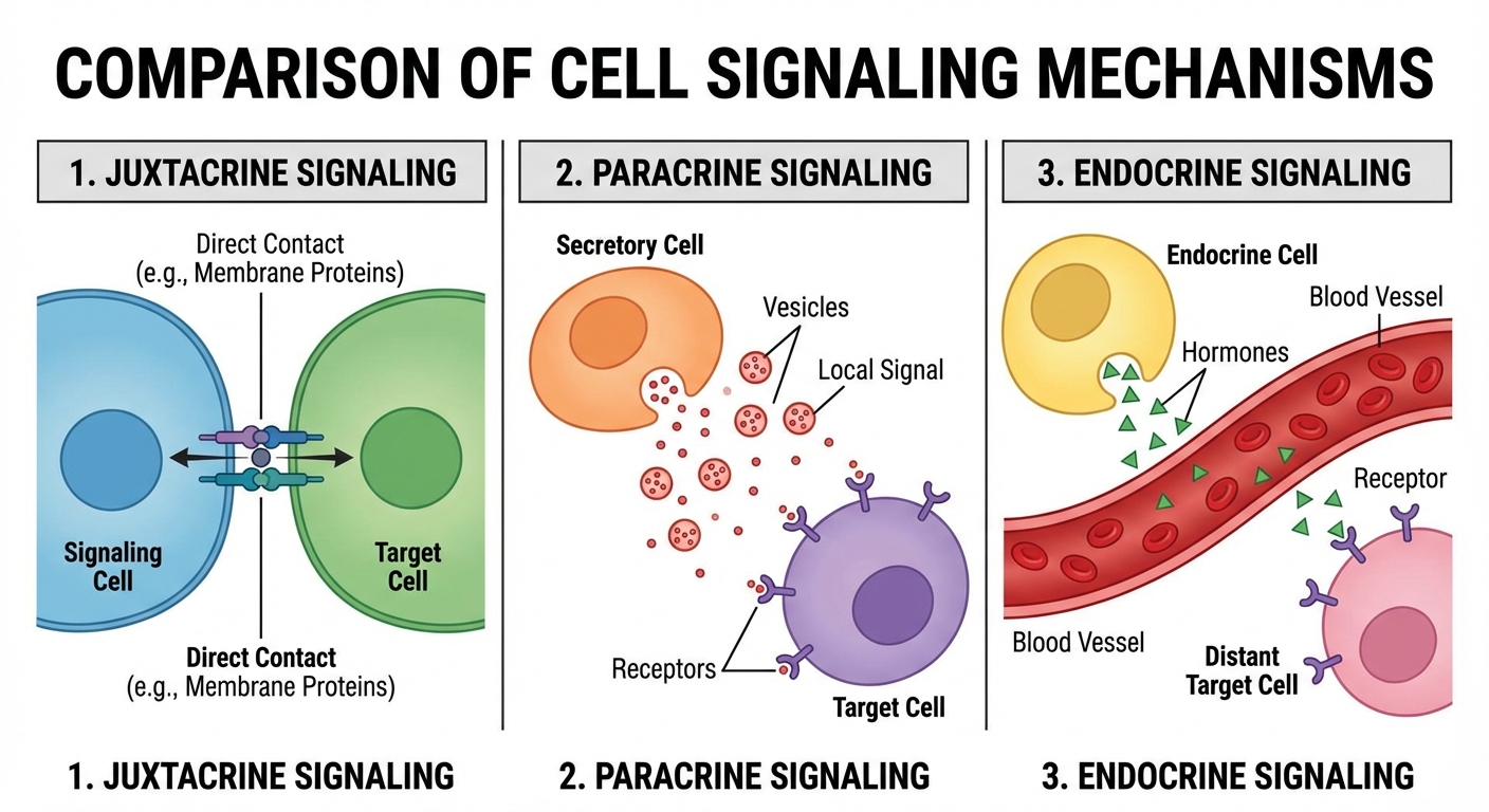
Cells communicate over varying distances. We classify these based on the proximity of the signaling cell to the target cell.
| Type | Distance | Description | Example |
|---|---|---|---|
| Juxtacrine | Contact | Direct contact between cells. | Plasmodesmata (plants), gap junctions (animals), immune cell interaction (APC to T-cell). |
| Paracrine | Short | Signals released into the extracellular fluid to nearby target cells. | Growth factors, synaptic signaling (neurotransmitters). |
| Endocrine | Long | Signals travel through the bloodstream (circulatory system) to distant targets. | Hormones like Insulin or Adrenaline. |
Introduction to Signal Transduction
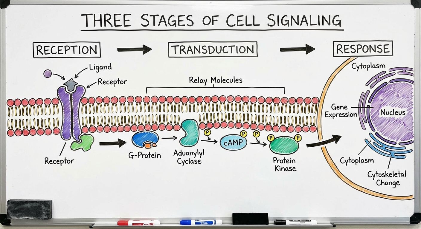
The Signal Transduction Pathway is the mechanism by which a signal on a cell's surface is converted into a specific cellular response. According to the College Board curriculum, this process occurs in three defined stages:
- Reception: The target cell detects a signaling molecule coming from outside the cell.
- Transduction: The conversion of the signal to a form that can bring about a specific cellular response.
- Response: The specific cellular effect brought about by the transduced signal.
1. Reception
Reception begins when a ligand binds to a receptor protein. This binding is highly specific—think of a lock and key.
- Conformational Change: The immediate result of ligand binding is a change in the shape of the receptor protein. This shape change is often the initial "transduction" of the signal.
- Intracellular vs. Membrane Receptors:
- Plasma Membrane Receptors: Bind to hydrophilic (water-soluble, polar) ligands that cannot cross the membrane (e.g., Epinephrine, Insulin).
- Intracellular Receptors: Found in the cytoplasm or nucleus. They bind to hydrophobic (lipid-soluble, nonpolar) ligands that can cross the membrane (e.g., Steroid hormones like Testosterone, Nitric Oxide).
Signal Transduction
Once the receptor is activated, the signal must be relayed through the cell. This stage usually involves a sequence of changes in a series of different molecules, known as a signal transduction pathway.
Protein Phosphorylation and Dephosphorylation
Many pathways transmit signals through a cascade of protein phosphorylation. This acts like a molecular "on/off" switch.
- Protein Kinase: An enzyme that transfers phosphate groups from ATP to a protein. This usually activates the protein.
- Protein Phosphatase: An enzyme that removes phosphate groups from proteins (dephosphorylation). This usually deactivates the protein, resetting the pathway.
Phosphorylation Cascade: A series of kinases that phosphorylate each other in turn. This allows for fine-tuning and amplification of the response.
Second Messengers
Not all components of a pathway are proteins. Second messengers are small, non-protein, water-soluble molecules or ions that spread throughout a cell by diffusion. They help relay the signal from the membrane to the interior.
- Cyclic AMP (cAMP):
- Adenylyl cyclase (enzyme) converts ATP to cAMP in response to a signal (like Epinephrine).
- cAMP usually activates Protein Kinase A (PKA), which phosphorylates other proteins.
- Calcium Ions ($Ca^{2+}$):
- Cells maintain very low cytosolic $Ca^{2+}$ concentrations.
- Signaling causes an influx of $Ca^{2+}$ from the ER or outside the cell, triggering responses (e.g., muscle contraction, exocytosis).
Signal Amplification
One of the most important features of transduction is amplification. A single ligand binding to a receptor can trigger the production of thousands of second messengers, which activate millions of enzymes.
Example: 1 molecule of Epinephrine $\rightarrow$ $10^8$ molecules of Glucose released from Glycogen.
Changes in Signal Transduction Pathways
The environment and genetic makeup can alter how signaling pathways function. These alterations are frequently tested on the AP exam.
1. Mutations and Receptor Function
A mutation in the DNA encoding a receptor or a component enzyme can alter the protein's primary structure, leading to a change in tertiary shape.
- Loss of Function: If the receptor shape changes, the ligand may no longer bind, halting the pathway completely.
- Constitutive Activation: Some mutations cause a receptor or kinase (like Ras) to remain in the "active" state even without a ligand. This uncontrolled signaling is a hallmark of cancer.
2. Chemicals and Inhibitors
External chemicals can interfere with pathways:
- Inhibitors: Block the receptor site so the natural ligand cannot bind (competitive inhibition) or bind elsewhere to change the receptor shape (allosteric inhibition).
- Activators: Mimic the ligand and trigger the pathway artificially.
3. Feedback Mechanisms
Pathways are regulated by the physiological state of the organism.
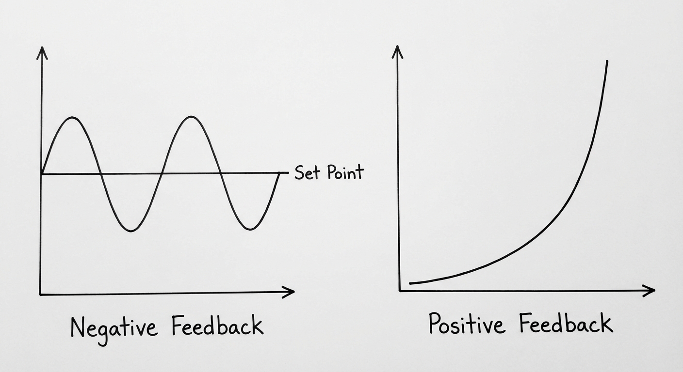
- Negative Feedback: The output of the pathway inhibits the upstream steps, returning the system to a set point (Homeostasis).
- Example: High ATP levels inhibit enzymes in Glycolysis.
- Positive Feedback: The output amplifies the initial stimulus, moving the system away from the set point.
- Example: Oxytocin during childbirth increases contractions, which causes more Oxytocin release.
Common Mistakes & Pitfalls
- The Ligand Entry Trap: Students often assume the signaling molecule enters the cell to do the work.
- Correction: Hydrophilic ligands (most common in exam questions) never enter the cell. They stay outside and poke the receptor. Only hydrophobic lipids (steroids) enter.
- Kinase vs. Phosphatase: Confusion over which enzyme does what.
- Mnemonic: Kinases Kindle the fire (turn things on/add phosphate). Phosphatase takes it away.
- Same Ligand, Different Response: Assuming one ligand always does the same thing.
- Correction: The response depends on the cell type and the specific intracellular proteins available. Epinephrine causes liver cells to break down glycogen but causes heart cells to beat faster.
- Stopping the Signal: Forgetting that pathways must be shut off.
- Correction: If the ligand detaches, or if GTP hydrolyzes to GDP, or if phosphatases act, the signal stops. Perpetual signaling usually indicates disease/toxins (e.g., Cholera toxin freezes a G-protein in the active state).