Mastering the Microscopic: Cell Structure, Function, and Transport
Unit Overview: The Fundamental Unit of Life
Unit 2 of the AP Biology curriculum moves from the chemistry of life to the physical containers of life: cells. This unit focuses on how cells maintain an internal environment that is different from their external environment, how structure dictates function (a recurring theme), and the evolutionary history of eukaryotic cells.
Cell Size and Exchange Efficiency
The physical size of a cell is limited by the laws of physics and geometry. Cells must efficiently exchange nutrients and wastes with their environment to sustain metabolism.
Surface Area-to-Volume Ratio (SA:V)
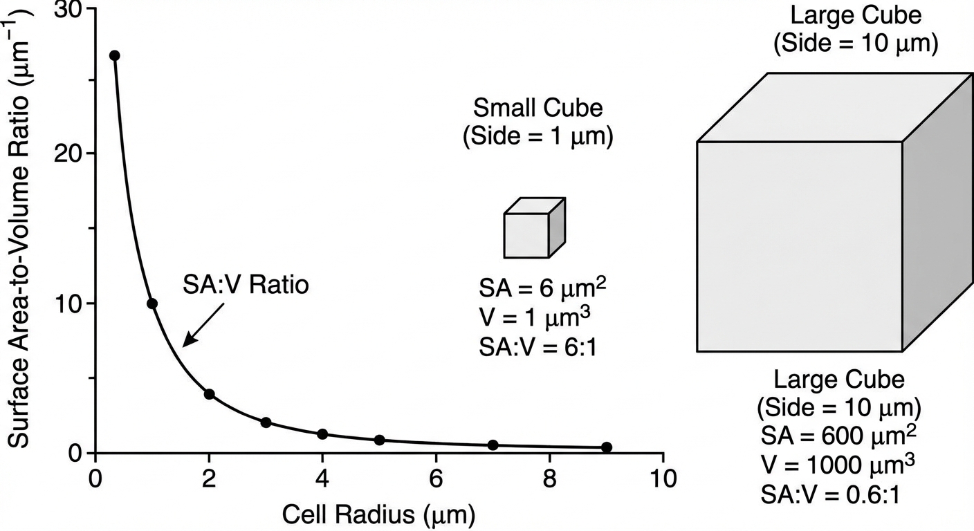
- Concept: The surface area represents the plasma membrane (where exchange happens), while the volume represents the cytoplasm/organelles (where metabolic activity happens).
- The Rule: As a cell grows larger, its volume increases much faster than its surface area. Therefore, smaller cells have a higher SA:V ratio, making them more efficient at material exchange.
Mathematical Relationship
Comparing a small cube ($side = 1$) to a large cube ($side = 5$):
- Small Cube: $SA = 6$, $Vol = 1$. Ratio $\approx 6:1$
- Large Cube: $SA = 150$, $Vol = 125$. Ratio $\approx 1.2:1$
The small cell has 6 units of membrane for every 1 unit of volume to service. The large cell struggles to import enough food to support its massive volume.
Adaptations to Increase Surface Area
Organisms have evolved specialized structures to maximize surface area without increasing volume significantly:
- Root Hairs: Extensions on plant roots to maximize water absorption.
- Villi/Microvilli: Finger-like projections in the human small intestine to maximize nutrient absorption.
- Elephant Ears: Large, flat surface areas to dissipate heat (thermal exchange).
Subcellular Components & Compartmentalization
Life is categorized into Prokaryotes (Domains Bacteria and Archaea) and Eukaryotes (Domain Eukarya). While they share common ancestry, their structural complexity differs.
Prokaryotic vs. Eukaryotic Cells
| Feature | Prokaryotes (Bacteria/Archaea) | Eukaryotes (Plants/Animals/Fungi) |
|---|---|---|
| Nucleus | No (DNA in nucleoid region) | Yes (Membrane-bound) |
| Membrane-bound Organelles | No | Yes (Mitochondria, Lysosomes, etc.) |
| DNA Shape | Circular, single chromosome | Linear, multiple chromosomes |
| Size | Small (1-5 μm) | Large (10-100 μm) |
| Ribosomes | Yes (Smaller, 70S) | Yes (Larger, 80S) |
The Importance of Compartmentalization
Eukaryotic cells use internal membranes to partition the cell into specialized regions. This allows:
- Enzymatic isolation: Incompatible chemical reactions can occur simultaneously (e.g., lysosomes digest materials at acidic pH while the cytosol remains neutral).
- Efficiency: Enzymes and substrates are concentrated in specific areas rather than diffused throughout the whole cell.
Organelle Function Breakdown
1. Genetic Control
- Nucleus: Contains DNA (chromatin) and the nucleolus (site of rRNA synthesis and ribosome assembly). Surrounded by a double membrane (nuclear envelope).
- Ribosomes: The sites of translation (protein synthesis). Not membrane-bound.
- Free Ribosomes: Float in cytosol; make proteins for use within the cell.
- Bound Ribosomes: Attached to Rough ER; make proteins for export or membrane insertion.
2. The Endomembrane System (Manufacturing & Shipping)
- Rough Endoplasmic Reticulum (RER): Studded with ribosomes. Compartmentalizes the cell and provides mechanical support. Synthesizes glycoproteins.
- Smooth Endoplasmic Reticulum (SER): Lacks ribosomes. Functions: Lipid synthesis, detoxification of poisons (liver), and calcium storage (muscles).
- Golgi Complex: Series of flattened membrane sacs (cisternae). It modifies, sorts, and packages proteins entering from the ER. Think of it as the UPS center of the cell.
3. Energy Transformers
- Mitochondria: Site of cellular respiration (ATP production).
- Structure: Double membrane. The inner membrane is highly folded (cristae) to increase surface area for electron transport chain enzymes. The fluid center is the matrix (Krebs cycle location).
- Chloroplasts: Site of photosynthesis (Plants/Algae).
- Structure: Double membrane. Contains stacks of discs called thylakoids (light reactions) within a fluid called stroma (Calvin cycle).
4. Waste and Storage
- Lysosomes: Membrane sacs containing hydrolytic enzymes. used for intracellular digestion, recycling cell parts (autophagy), and apoptosis (programmed cell death).
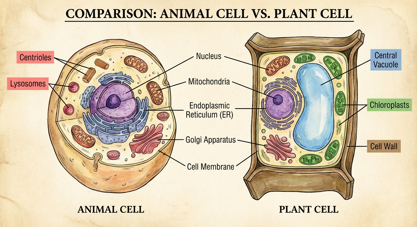
- Vacuoles: Large sacs for storage. Plants have a large Central Vacuole that stores water and maintains turgor pressure against the cell wall.
The Plasma Membrane
The Fluid Mosaic Model
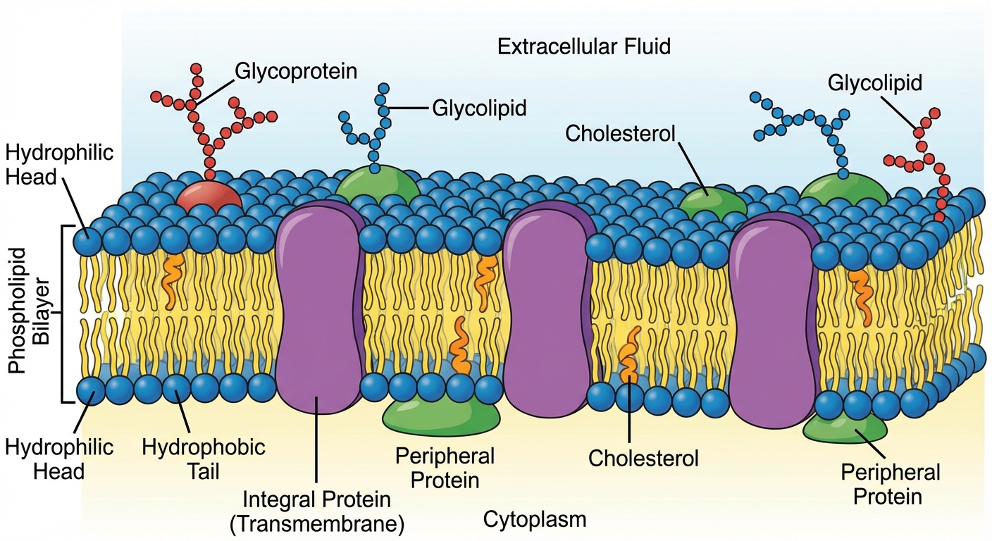
The membrane is a dynamic, moving mosaic of lipids and proteins.
- Phospholipids: The foundation. They are amphipathic (hydrophilic head, hydrophobic tails). They spontaneously form a bilayer where tails face inward (away from water) and heads face outward.
- Proteins:
- Integral: Embedded in the bilayer; usually amphipathic.
- Peripheral: Loosely attached to the surface.
- Cholesterol: Modulates fluidity. At high temps, it restrains movement (prevents melting). At low temps, it prevents packing (prevents freezing).
- Carbohydrates: Glycolipids and Glycoproteins act as ID tags for cell-to-cell recognition.
Membrane Permeability & Transport
Selective Permeability
The structure of the lipid bilayer dictates what can pass through:
- Easy Pass: Small, nonpolar molecules ($N2$, $O2$, $CO_2$) pass freely via simple diffusion.
- Hard Pass: Hydrophilic substances (large polar molecules like glucose, or ions like $Na^+$, $K^+$) are repelled by the hydrophobic core. They require transport proteins.
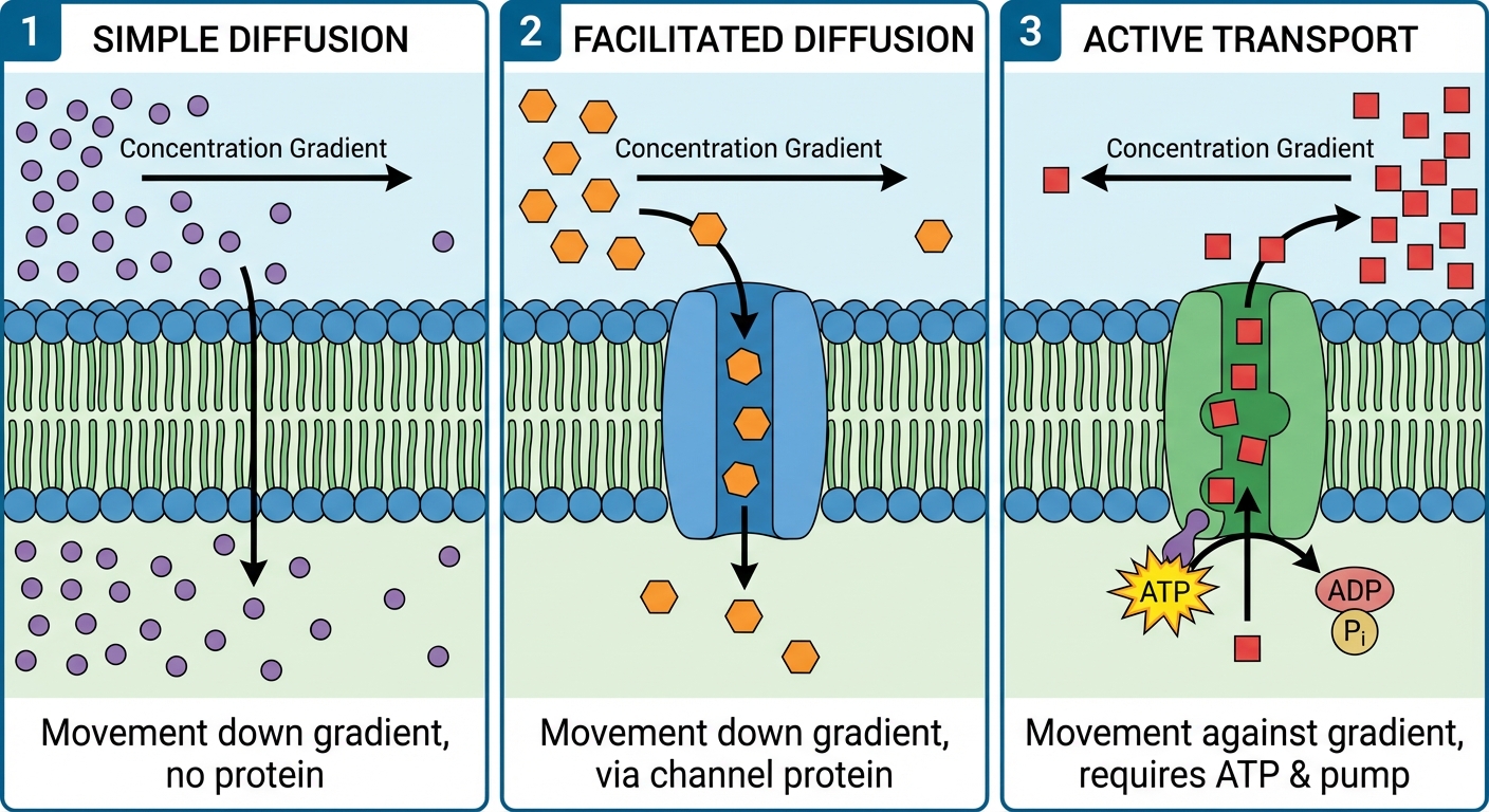
1. Passive Transport (No Energy Required)
Movement down the concentration gradient (High $\rightarrow$ Low).
- Simple Diffusion: Movement of nonpolar molecules directly through the bilayer.
- Facilitated Diffusion: Movement of polar molecules/ions through transport proteins.
- Channel Proteins: Hydrophilic tunnels (e.g., Aquaporins for water).
- Carrier Proteins: Change shape to shuttle molecules across.
2. Active Transport (Requires ATP)
Movement against the concentration gradient (Low $\rightarrow$ High).
- Pumps: Proteins that force molecules across.
- Example: Sodium-Potassium Pump ($Na^+/K^+$ ATP-ase). Pumps 3 $Na^+$ OUT and 2 $K^+$ IN. Essential for nerve transmission.
- Cotransport: Uses the energy stored in an electrochemical gradient (generated by a pump) to move another substance.
- Example: Sucrose-$H^+$ cotransporter in plants.
3. Bulk Transport (Endocytosis & Exocytosis)
Used for macromolecules too large for protein channels. Requires energy.
- Exocytosis: Internal vesicles fuse with the membrane to release contents (e.g., neurons releasing neurotransmitters).
- Endocytosis: Cell takes in matter by forming new vesicles.
- Phagocytosis: "Cell eating" (solids).
- Pinocytosis: "Cell drinking" (fluids).
- Receptor-Mediated: Specific uptake triggered by ligand binding.
Tonicity and Osmoregulation
Osmosis is the diffusion of water across a semipermeable membrane. Water moves from High Water Potential $\rightarrow$ Low Water Potential.
Tonicity Types
| Solution Type | Solute Concentration | Water Movement | Animal Cell | Plant Cell |
|---|---|---|---|---|
| Hypotonic | Lower outside | Into Cell | Lyses (Bursts) | Turgid (Normal/Healthy) |
| Isotonic | Equal | Net zero | Normal | Flaccid |
| Hypertonic | Higher outside | Out of Cell | Shriveled | Plasmolyzed (Wilts) |
Water Potential ($\Psi$)
A measure of the potential energy of water. Water flows from high $\Psi$ to low $\Psi$.
Formula:
- $\Psi_P$ (Pressure Potential): Physical pressure (turgor). Open beaker = 0.
- $\Psi_S$ (Solute Potential): Always zero (pure water) or negative. Adding solute lowers water potential.
Solute Potential Formula:
- $i$ = Ionization constant (1 for sucrose, 2 for NaCl)
- $C$ = Molar concentration
- $R$ = Pressure constant (0.0831 liter bars/mole K)
- $T$ = Temperature in Kelvin ($^\circ C + 273$)
Worked Example:
A cell with $\PsiP = 1.0$ bar and $\PsiS = -3.5$ bars is placed in a solution with $\PsiP = 0$ bar and $\PsiS = -4.0$ bars.
- $\Psi_{cell} = 1.0 + (-3.5) = -2.5$ bars
- $\Psi_{solution} = 0 + (-4.0) = -4.0$ bars
- Since $-2.5 > -4.0$, water moves from the cell into the solution. The cell will shrink.
Origins of Compartmentalization
Endosymbiotic Theory
This theory explains how eukaryotic cells evolved from prokaryotic ancestors. It states that an early ancestor of eukaryotic cells engulfed an oxygen-using non-photosynthetic prokaryotic cell. Eventually, the engulfed cell became an endosymbiont (a cell living within another cell).
Key Evidence (MDR):
- Membranes: Mitochondria and chloroplasts have double membranes (inner membrane similar to prokaryotes).
- DNA: They have their own circular DNA, distinct from the nucleus.
- Ribosomes: They have their own ribosomes that are similar in size to prokaryotic ribosomes (70S).
- Reproduction: They reproduce independently via binary fission.
Common Mistakes & Pitfalls
- Diffusion Misconception: Students often think molecules stop moving in an isotonic solution. Correction: Molecules continue to move, but there is no net change in concentration.
- Cell Wall vs. Membrane: Don't confuse them. All cells have membranes; NOT all cells have walls. The wall is for structure (plants/fungi/bacteria), the membrane is for transport control.
- Solute Potential Math: Remember the negative sign! The more solute you add, the lower (more negative) the potential becomes. A -10 bar solution has lower potential than a -5 bar solution.
- Hypotonic/Hypertonic: These are relative terms. Solution A is only hypertonic compared to Solution B. Always identify the frame of reference.
- Excretion vs Secretion: Secretion is the release of a useful product (enzyme/hormone) via exocytosis. Excretion is waste removal.