AP Biology Unit 2: Cell Structure and Organization
AP Biology Unit 2: Cell Structure and Organization
This unit focuses on the fundamental unit of life: the cell. For the AP exam, you must understand not just what the parts of the cell are, but how their specific structures allow them to perform their functions efficiently. This relationship between structure and function is a recurring theme.
2.1 & 2.2 Cell Structure: Subcellular Components and Function
Cells are organized systems where specific sub-components (organelles) facilitate specific metabolic processes.
Ribosomes
Ribosomes are the cellular machinery responsible for protein synthesis. They are not membrane-bound and are found in all known forms of life (prokaryotes and eukaryotes), reflecting the common ancestry of all living things.
- Structure: Composed of ribosomal RNA (rRNA) and protein. Consists of a large and a small subunit.
- Locations:
- Free Ribosomes: Float in the cytosol; synthesize proteins used within the cytosol.
- Bound Ribosomes: Attached to the Rough ER; synthesize proteins for membrane insertion, packaging within organelles, or secretion (export).
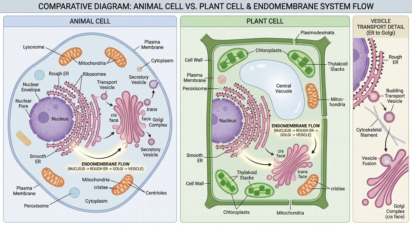
The Endomembrane System
This is a group of membranes and organelles in eukaryotic cells that work together to modify, package, and transport lipids and proteins.
1. Endoplasmic Reticulum (ER)
The ER is a network of membrane tubes within the cytoplasm. The structure differentiates its function:
- Rough ER:
- Structure: Surface is studded with ribosomes.
- Function: Compartmentalizes the cell; provides structural support; synthesizes proteins intended for the cell membrane, lysosomes, or export.
- Smooth ER:
- Structure: No ribosomes attached.
- Function: Synthesizes lipids (membrane phospholipids), metabolizes carbohydrates, and detoxifies drugs and poisons (abundant in liver cells).
2. The Golgi Complex (Golgi Apparatus)
- Structure: A series of flattened membrane sacs called cisternae.
- Function: Receives transport vesicles from the ER, modifies the proteins (e.g., glycosylation—adding sugar chains), folds them, and packages them into new vesicles for sorting.
- Cis face: Receives vesicles.
- Trans face: Ships vesicles out.
3. Lysosomes
- Structure: Membrane-enclosed sacs containing hydrolytic enzymes.
- Function: Intracellular digestion.
- Recycling organic materials (autophagy).
- Programmed cell death (apoptosis).
- Important: The interior is acidic; if the lysosome breaks, the enzymes function poorly in the neutral cytosol, preventing accidental self-digestion of the cell.
4. Vacuoles
- Structure: Membrane-bound sacs.
- Function: Storage and release of macromolecules and waste.
- Central Vacuole (Plants): Maintains turgor pressure against the cell wall to keep the plant upright; stores water and ions.
- Contractile Vacuole (Protists): Pumps out excess water to prevent bursting (osmoregulation).
Energy-Capturing Organelles
1. Mitochondria
The site of cellular respiration (ATP production). Found in nearly all eukaryotic cells.
- Double Membrane:
- Outer membrane: Smooth.
- Inner membrane: Highly folded into cristae to increase surface area for the Electron Transport Chain (ETC).
- Matrix: The fluid-filled inner space where the Krebs Cycle (Citric Acid Cycle) occurs.
2. Chloroplasts
The site of photosynthesis. Found in plants and algae.
- Double Membrane: Outer and inner membranes.
- Thylakoids: Membranous sacs stacked into grana. The light-dependent reactions occur here (chlorophyll pigments reside in these membranes).
- Stroma: The fluid surrounding the thylakoids; site of the Calvin Cycle (carbon fixation).
| Feature | Mitochondria | Chloroplasts |
|---|---|---|
| Primary Function | Cellular Respiration (ATP generation) | Photosynthesis (Sugar generation) |
| Key Internal Structure | Cristae (folded inner membrane) | Thylakoids (stacked sacs) |
| Fluid Compartment | Matrix | Stroma |
| Found in | Almost all Eukaryotes | Plants & Algae |
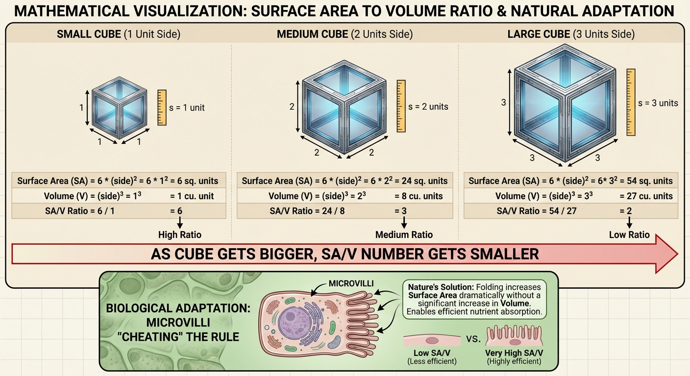
2.3 Cell Size
Cells must be small to efficiently exchange materials with their environment. The movement of waste out of the cell and nutrients into the cell depends on the Surface Area-to-Volume Ratio ($SA/V$).
The Math of SA/V
As a cell increases in size, its volume ($V$) increases much faster than its surface area ($SA$).
- Volume dictates the amount of metabolic activity (how much nutrient is needed/waste produced).
- Surface Area dictates the rate of exchange (how fast things can get in/out).
SA{cube} = 6s^2 V{cube} = s^3
As the side length ($s$) increases, the ratio $\frac{6s^2}{s^3} = \frac{6}{s}$ decreases.
Biological Implications
- High SA/V Ratio: Smaller cells have a higher ratio, allowing for efficient exchange. This helps facilitate thermal energy dissipation and gas exchange.
- Limitations: If a cell grows too large, the plasma membrane cannot transport enough O$_2$ or nutrients to feed the massive volume inside. The cell must divide or die.
Adaptations to Increase Surface Area
Cells that need to maximize exchange without being microscopic utilize structural modifications:
- Root Hairs: Long, thin projections on plant roots increase absorption of water/minerals.
- Villi/Microvilli: Finger-like projections in the small intestine lining increase absorption of nutrients.
- Flattening: Elephant ear cells are flat and thin to dissipate heat.
2.10 Cell Compartmentalization
Compartmentalization refers to the presence of membrane-bound organelles that separate different metabolic processes within the same cell.
Benefits of Compartmentalization
- Minimize Competing Reactions: Incompatible reactions can occur simultaneously. (e.g., synthesis of fatty acids in one area vs. breakdown of fatty acids in another).
- Increased Efficiency: Enzymes and substrates are concentrated in specific areas (e.g., Krebs cycle enzymes are concentrated in the mitochondrial matrix).
- Specific Environments: Organelles maintain internal conditions distinct from the cytosol (e.g., Lysosomes maintain a pH of ~5, while cytosol is ~7.2).
Prokaryotes vs. Eukaryotes
- Eukaryotes: Extensive internal membrane systems and membrane-bound organelles.
- Prokaryotes: Generally lack internal membrane-bound organelles. However, they have specialized internal regions (e.g., nucleoid region) and some perform metabolic functions on infoldings of the plasma membrane.
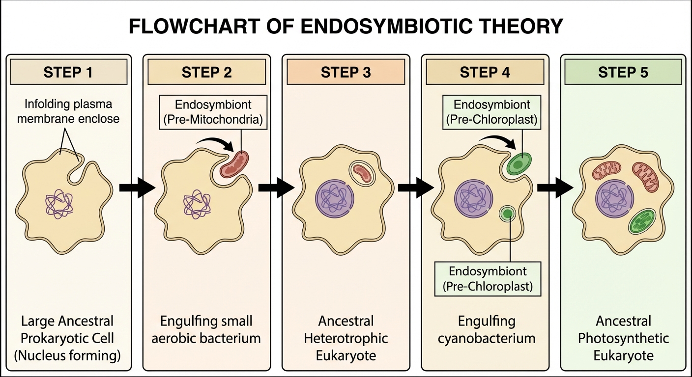
2.11 Origins of Cell Compartmentalization
Where did eukaryotic cells come from? The leading explanation is the Endosymbiotic Theory.
The Endosymbiotic Theory
This theory states that membrane-bound organelles (specifically mitochondria and chloroplasts) evolved from previously free-living prokaryotic cells that were engulfed by a larger ancestral prokaryote (or early eukaryote) via phagocytosis.
The Narrative:
- An ancestral anaerobe engulfed an aerobic bacterium.
- Instead of digesting it, a symbiotic relationship formed: the host provided protection/nutrients, and the engulfed bacterium provided ATP.
- This engulfed bacterium evolved into the mitochondrion.
- Later, a descendant of this cell engulfed a photosynthetic bacterium (cyanobacterium), which evolved into the chloroplast.
Evidence for Endosymbiosis (The "DR. D" Mnemonic)
To confirm this theory, scientists look at how mitochondria/chloroplasts resemble bacteria:
- D - DNA: Mitochondria and chloroplasts have their own circular, naked DNA (like bacteria), separate from the nucleus.
- R - Ribosomes: They contain 70S ribosomes (prokaryotic size), whereas the eukaryotic cytoplasm has 80S ribosomes.
- D - Double Membrane: The inner membrane belongs to the original bacterium; the outer membrane is formed from the host's vesicle during engulfment.
- Reproduction: They replicate independently of the cell via binary fission, just like bacteria.
Common Mistakes & Pitfalls
Confusion over Cell Walls vs. Membranes:
- Mistake: Thinking only plants have cell walls and only animals have cell membranes.
- Correction: All cells (prokaryotic and eukaryotic) have a cell membrane (plasma membrane). Plants, fungi, and most prokaryotes also have a cell wall.
Misunderstanding SA/V Ratio:
- Mistake: Thinking a "larger" ratio means a larger cell.
- Correction: Large SA/V ratio = Small Cell (or highly folded). Small SA/V ratio = Large/Inefficient Cell.
Protein Path:
- Mistake: Thinking proteins go from Smooth ER → Golgi.
- Correction: Proteins for secretion consist of: Ribosome → Rough ER → Transport Vesicle → Golgi → Secretory Vesicle → Plasma Membrane.
Plant Cell Mitochondria:
- Mistake: Thinking plants have chloroplasts instead of mitochondria.
- Correction: Plant cells have both. They need chloroplasts to make sugar and mitochondria to break that sugar down into ATP.