AP Biology: Unit 2 Complete Guide
Unit 2: Cell Structure and Function
Unit Overview
In AP Biology Unit 2, the central theme is the inextricable link between a cell's physical form and its biological function. You will move beyond simply naming organelles to understanding how their specific structures (like membrane folding) allow for life-sustaining processes. This unit covers the evolutionary origins of eukaryotes (Endosymbiosis), the physics of membrane transport, and the mathematical modeling of water movement (Water Potential).
2.1 & 2.2 Subcellular Components
Compartmentalization is the defining characteristic of eukaryotic cells. Internal membranes create separate micro-environments (e.g., specific pH or ion concentrations) that allow incompatible metabolic processes to occur simultaneously within the same cell.
The Genetic & Protein Machinery
The Nucleus
- Function: Houses the genome (DNA) and directs protein synthesis via mRNA.
- Structure: Surrounded by the Nuclear Envelope (double membrane) with pores that regulate traffic (mRNA out, proteins in).
- Nucleolus: A dense region inside the nucleus responsible for synthesis of rRNA and assembly of ribosome subunits.
Ribosomes (The Universal Organelle)
- Definition: Non-membrane-bound complexes of rRNA and protein that perform Translation (protein synthesis).
- Evolutionary Significance: Found in all domains of life (Bacteria, Archaea, Eukarya), serving as primary evidence of common ancestry.
- Location = Function:
- Free Ribosomes: Suspended in cytosol. Produce proteins that function within the cytosol (e.g., enzymes for glycolysis).
- Bound Ribosomes: Attached to the Rough ER. Produce proteins aimed for secretion, the plasma membrane, or lysosomes.
The Endomembrane System
This group of membranes regulates protein traffic and performs metabolic functions. The pathway generally flows: Nucleus $\rightarrow$ Rough ER $\rightarrow$ Transport Vesicle $\rightarrow$ Golgi $\rightarrow$ Plasma Membrane/Lysosome.
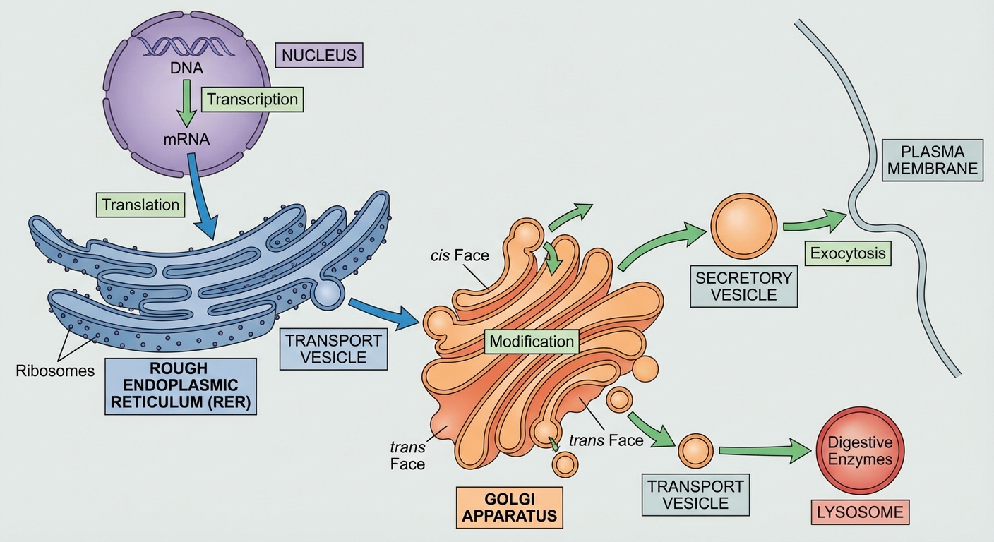
Endoplasmic Reticulum (ER)
- Rough ER: Studded with bound ribosomes. Compartmentalizes the cell and modifies proteins (e.g., folding) destined for export.
- Smooth ER: Lacks ribosomes. Functions include lipid synthesis, detoxification of poisons (liver cells), and calcium ion storage (muscle cells).
Golgi Complex (The Post Office)
- Structure: Flattened membrane sacs called cisternae.
- Directionality:
- Cis face: Receives vesicles from the ER.
- Trans face: Ships vesicles out.
- Function: Corrects folding and chemically modifies proteins. A vital modification is Glycosylation (adding carbohydrate tags), treating proteins like barcoded packages for sorting.
Lysosomes (Animal Cells)
- Function: Intracellular digestion using hydrolytic enzymes. These enzymes function best in acidic environments ($pH \approx 5$).
- Phagocytosis: Digestion of food or pathogens engulfed by the cell.
- Autophagy: Breaks down its own damaged organelles for recycling.
- Apoptosis: Programmed cell death (e.g., removing webbing between fingers in embryos).
Vacuoles
- Food Vacuoles: Formed via phagocytosis.
- Contractile Vacuoles: Pump excess water out of freshwater protists to prevent bursting (osmoregulation).
- Large Central Vacuole (Plants): Stores water and ions. Creates Turgor Pressure against the cell wall, which is essential for plant structural support.
Energy Organelles
Both mitochondria and chloroplasts are double-membraned, support the Endosymbiotic Theory (see Section 2.11), and utilize high surface area to maximize energy production.
| Organelle | Function | Key Structural Feature | Significance of Structure |
|---|---|---|---|
| Mitochondria | Cellular Respiration (ATP Production) | Cristae (folds of inner membrane) | Maximizes surface area for the Electron Transport Chain (ETC) and ATP Synthase. |
| Chloroplast | Photosynthesis (Plants/Algae) | Thylakoids stacked into Grana | Increases surface area for photosystems (light-dependent reactions) to capture solar energy. |
2.3 Cell Size & Exchange
Biological systems are constrained by geometry. Cells must exchange materials with the environment (nutrients in, waste/heat out) to maintain homeostasis.
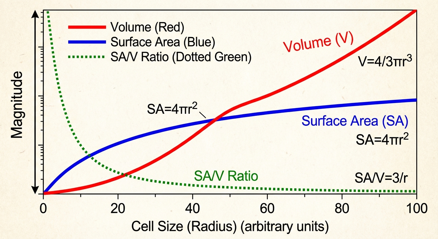
Surface Area to Volume Ratio ($SA:V$)
- Surface Area ($SA$): Supply. Determines the rate at which material enters/leaves.
- Volume ($V$): Demand. Determines metabolic rate (how much food is needed/waste produced).
The Rule: Cells require a High SA:V Ratio. As a cell increases in size, its volume ($s^3$) grows much faster than its surface area ($s^2$). If a cell becomes too large, the plasma membrane cannot facilitate enough exchange to support the metabolic needs of the cytosol.
Mathematical Proof
Consider a cube with side length $s$:
- Small Cell ($s=1$): $Ratio = 6:1$
- Large Cell ($s=5$): $Ratio = 1.2:1$
- Conclusion: The smaller cell is significantly more efficient at exchange.
Structural Adaptations
To bypass surface area limitations, large cells or specialized tissues evolve specific shapes:
- Membrane Folding: Inner mitochondrial membranes (cristae).
- Projections: Microvilli on intestinal cells and Root Hairs on plants effectively increase $SA$ without significantly increasing $V$.
- Flattening: Fish gills or elephant ears (for heat exchange).
2.4 & 2.5 Plasma Membrane Structure
The membrane is defined by the Fluid Mosaic Model: a fluid lipid bilayer embedded with a mosaic of proteins, steroids, and carbohydrates.
Components
Phospholipids (The Barrier)
- Amphipathic: Possess both a hydrophilic head (polar/phosphate) and a hydrophobic tail (nonpolar/fatty acid).
- Arrangement: Spontaneously form a bilayer with tails hiding from water and heads facing the aqueous cytosol/extracellular fluid.
Membrane Proteins
- Integral: Span the membrane (transmembrane). Hydrophobic regions sit within the lipid tails; hydrophilic regions extend out.
- Peripheral: Loosely bound to the surface.
- Functions: Transport, signal transduction, enzymatic activity, and cell-cell recognition.
Cholesterol
- A steroid wedged between phospholipids.
- Function: Regulates membrane fluidity. At high temps, it stabilizes the membrane (prevents melting). At low temps, it prevents phospholipids from packing too tightly (prevents freezing).
Carbohydrates (Glycoproteins/Glycolipids)
- Act as identification markers (e.g., blood types).
Selective Permeability
The hydrophobic core regulates entry.
- Easy Passage: Small, nonpolar molecules ($N2, O2, CO_2$) diffuse freely.
- Difficult/Blocked: Large polar molecules (glucose) and ions ($Na^+, K^+, Cl^-$) are repelled by the hydrophobic core and require transport proteins.
- Water: Small but polar. Moves very slowly naturally; requires Aquaporins for rapid transport.
Cell Walls (Not Just Plants)
Cell walls provide structural support and permeability barriers, but they are extracellular structures (outside the membrane).
- Plants: Cellulose.
- Fungi: Chitin.
- Prokaryotes: Peptidoglycan.
2.6 - 2.9 Transport Mechanisms
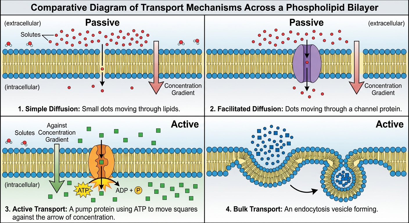
Passive Transport
Movement down the concentration gradient (High $\rightarrow$ Low). No ATP required.
- Simple Diffusion: Nonpolar molecules slide directly through the phospholipids.
- Osmosis: The diffusion of water across a semipermeable membrane from High Water Potential to Low Water Potential.
- Facilitated Diffusion: Movement of polar/charged molecules via proteins.
- Channel Proteins: Hydrophilic tunnels (e.g., Aquaporins).
- Carrier Proteins: Change shape to shuttle molecules (e.g., Glucose transporter).
Active Transport
Movement against the concentration gradient (Low $\rightarrow$ High). Requires energy (ATP).
- Protein Pumps: Maintain electrochemical gradients.
- $Na^+/K^+$ Pump: (Animal cells) Pumps 3 $Na^+$ out and 2 $K^+$ in. Creates a charge difference essential for nerve signaling.
- Proton Pump: (Plants/Fungi/Bacteria) Pumps $H^+$ out to create a gradient for cotransport.
- Cotransport: Uses the energy stored in an electrochemical gradient (generated by a pump) to move a second molecule against its gradient.
- Example: Plants use the $H^+$ gradient to drag Sucrose into the cell.
Bulk Transport
Transport of large molecules via vesicles (Requires ATP).
- Exocytosis: Internal vesicles fuse with the membrane to secrete contents (hormones, neurotransmitters).
- Endocytosis: Membrane pinches in to take up material.
- Phagocytosis: Cell eating (solids).
- Pinocytosis: Cell drinking (extracellular fluid).
- Receptor-Mediated Endocytosis: Specific uptake via ligand binding (e.g., LDL cholesterol).
2.8 Osmoregulation & Water Potential
Key Rule: Water always moves from High Water Potential to Low Water Potential.
Tonicity
Tonicity describes the environment relative to the cell.
| Solution Type | Solute Concentration | Water Movement | Plant Cell Status | Animal Cell Status |
|---|---|---|---|---|
| Hypotonic | Lower outside | Into Cell | Turgid (Normal) | Lysed (Bursts) |
| Isotonic | Equal | Net movement zero | Flaccid | Normal |
| Hypertonic | Higher outside | Out of Cell | Plasmolyzed | Shriveled |
Water Potential Math
The general formula:
- $\Psi_p$ (Pressure Potential): Physical pressure from the cell wall.
- Open beaker = $0$.
- Turgid plant cell = Positive (+).
- $\Psi_s$ (Solute Potential): Effect of solute concentration.
- Pure water = $0$.
- Any solution = Negative (adding solute lowers potential).
Calculating Solute Potential
- $i$: Ionization Constant. (Sugar $= 1$, NaCl $= 2$).
- $C$: Molar Concentration.
- $R$: Pressure Constant ($0.0831$ L bars / mol K).
- $T$: Temperature in Kelvin (Celsius + 273).
Worked Example
Problem: A plant cell with a pressure potential ($\Psi_p$) of $+2$ bars is placed in a solution of $0.1M$ sucrose at $27^{\circ}C$. Where does the water move?
Solution:
- Calculate Cell Potential:
- Assume $\Psis$ of cell is unknown but $\Psi{cell}$ must be compared to solution.
- (Actually, usually the problem asks for equilibrium, let's look at the Solution Potential first).
- Calculate Solution $\Psi$:
- Open beaker $\rightarrow \Psi_p = 0$.
- Calculate $\Psi_s = -(1)(0.1)(0.0831)(300)$. (Note: $27C = 300K$)
- $\Psi_{solution} = -2.49$ bars.
- Comparison:
- If the cell's total $\Psi$ was higher than $-2.49$, water leaves the cell. If lower, water enters.
(Note: Always determine direction by comparing Total $\Psi$ of A vs Total $\Psi$ of B. High $\rightarrow$ Low).
2.10 & 2.11 Compartmentalization & Evolution
Compartmentalization
Membrane-bound organelles allow different parts of the cell to specialize. This increases efficiency by concentrating enzymes and substrates in one place while protecting the rest of the cell from dangerous byproducts (e.g., peroxisomes contain $H2O2$).
Endosymbiotic Theory
Explains the origin of eukaryotic cells. It hypothesizes that a large ancestor prokaryote engulfed a smaller, aerobic prokaryote (which became the mitochondrion) and later a photosynthetic prokaryote (which became the chloroplast).
Evidence (Mnemonic: DR. MAD)
- Division: Mitochondria/Chloroplasts replicate independently via binary fission.
- Ribosomes: They contain 70S ribosomes (bacteria-sized), not the 80S found in the eukaryotic cytosol.
- Membranes: They have double membranes (Inner = bacterial origin, Outer = host vesicle origin).
- Antibiotics: Their protein synthesis is inhibited by antibiotics that target bacteria.
- DNA: They possess their own circular, naked DNA.
Common Mistakes & Pitfalls
- Sign Errors in Water Potential: Students frequently forget that $-20$ is lower than $-5$. Water moves toward the more negative number (the hypertonic area). Think of it as water "falling" down to the lower number.
- Saturation Kinetics: Understanding that Facilitated Diffusion has a speed limit. Once all transport proteins are occupied (saturated), increasing the concentration gradient will not increase the rate of transport. Simple diffusion does not saturate.
- Active vs. Passive Confusion: Just because a protein is used does not mean it is active transport. Facilitated Diffusion uses proteins but is passive (High $\rightarrow$ Low). Active Transport requires ATP.
- Cell Wall vs Membrane: The cell wall is permeable to almost everything small. It does not select what enters the cell; the plasma membrane does that.
- Hypotonic Terminology: A solution is only "hypotonic" relative to a cell. You cannot just say "this water is hypotonic" without a point of comparison.