AP Biology Unit 2 Reference: Cell Structure & Function
Unit 2: Cell Structure and Function
Unit Overview
In AP Biology Unit 2, the central theme is the relationship between structure and function. Biology is not just about memorizing the names of organelles, but understanding how their specific physical attributes—such as the folding of the inner mitochondrial membrane or the amphipathic nature of phospholipids—dictate their efficiency and role within the cell. This unit covers the evolutionary history of eukaryotic cells (Endosymbiosis), how cells maintain homeostasis via the plasma membrane, and the mathematics of water potential.
2.1 & 2.2 Subcellular Components
Compartmentalization is the defining characteristic of eukaryotic cells. By using internal membranes, cells create separate local environments (e.g., maintaining an acidic pH in lysosomes while the cytosol remains neutral) where specific metabolic reactions can occur without interference or competing reactions.
The Nucleus and Ribosomes
Nucleus
- Function: Houses the cell's genetic sorting instructions (DNA). It is surrounded by a double membrane called the nuclear envelope with pores regulating entry and exit.
- Nucleolus: A dense region within the nucleus where rRNA is synthesized and ribosome subunits are assembled.
Ribosomes (The Protein Factory)
- Definition: Non-membrane bound structures responsible for Translation (protein synthesis).
- Structure: Composed of rRNA and protein. Consist of a large and small subunit.
- Evolutionary Marker: Found in all domains of life (Bacteria, Archaea, Eukarya), serving as evidence of common ancestry.
- Location determines Destination:
- Free Ribosomes: Floating in cytosol. Synthesize proteins that function within the cytosol (e.g., enzymes for glycolysis).
- Bound Ribosomes: Attached to the Rough ER. Synthesize proteins aimed for secretion, the plasma membrane, or lysosomes.
The Endomembrane System
This system regulates protein traffic and performs metabolic functions.
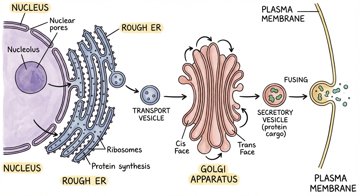
Endoplasmic Reticulum (ER)
- Rough ER: Studded with bound ribosomes. Compartmentalizes the cell and provides mechanical support. Major site of protein synthesis for export.
- Smooth ER: Lacks ribosomes. Functions in detoxification (abundant in liver cells), calcium storage (muscle cells), and lipid synthesis (membranes/hormones).
Golgi Complex (The Warehouse)
- Structure: Flattened membrane sacs called cisternae.
- cis face: Receives vesicles from the ER.
- trans face: Ships vesicles to the cytosol or membrane.
- Function: Modifies, folds, and packages proteins. A key modification is Glycosylation (adding carbohydrate tags) to create glycoproteins/glycolipids acting as molecular ID tags.
Lysosomes (Animal Cells)
- Definition: Membrane sacs containing hydrolytic enzymes.
- Function: Intracellular digestion.
- Phagocytosis: Digestion of pathogens or food particles.
- Autophagy: Recycling damaged organelles.
- Apoptosis: Programmed cell death (critical for embryonic development).
Vacuoles
- Food Vacuoles: Formed by phagocytosis.
- Contractile Vacuoles: Pump excess water out of freshwater protists (osmoregulation).
- Central Vacuole (Plants): Large storage for water and ions. Creates turgor pressure against the cell wall to maintain structural rigidity.
Energy Organelles
Both organelles support the Endosymbiotic Theory (see Section 2.11) and rely on surface area for efficiency.
Mitochondria
- Function: Cellular Respiration (ATP production).
- Structure: Double membrane. The Outer membrane is smooth; the Inner membrane is highly folded into Cristae.
- Significance: The folds (cristae) maximize Surface Area for the Electron Transport Chain (ETC) and ATP synthase. More surface area = More ATP.
- Matrix: The fluid-filled center where the Krebs Cycle occurs.
Chloroplasts
- Function: Photosynthesis (converting solar energy to chemical energy).
- Structure:
- Thylakoids: Discs containing chlorophyll and photosystems (Light Dep. Reactions).
- Grana: Stacks of thylakoids (increases Surface Area).
- Stroma: Fluid surrounding thylakoids (Calvin Cycle).
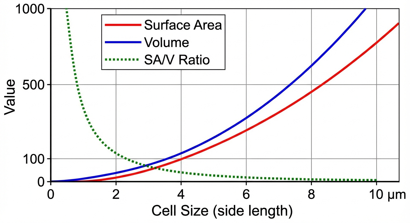
2.3 Cell Size & Exchange
The movement of resources (waste, nutrients, thermal energy) is constrained by geometry.
Surface Area to Volume Ratio ($SA:V$)
- Volume ($V$): Determines metabolic demand (how much food is needed/waste produced).
- Surface Area ($SA$): Determines the rate of exchange (how fast food enters/waste leaves).
The Rule: Cells must maintain a High Surface Area-to-Volume Ratio to survive. As a cell grows, Volume increases ($s^3$) much faster than Surface Area ($s^2$). If a cell gets too big, it cannot transport nutrients fast enough to service its volume, and it dies.
Formulas
For a cube with side $s$:
Adaptations for Exchange
Large/complex cells utilize structural modifications to increase SA without significantly increasing V:
- Root Hairs: Extensions on plant roots maximize atomic uptake.
- Microvilli: Finger-like projections on intestinal cells maximize nutrient absorption.
- Membrane Folding: The inner membranes of mitochondria and chloroplasts.
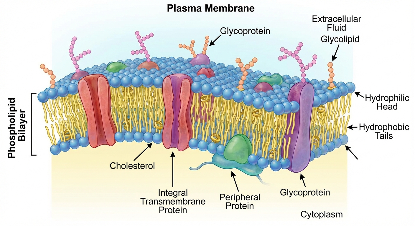
2.4 & 2.5 Plasma Membrane Structure
The membrane is described by the Fluid Mosaic Model—a moving sea of lipids with a mosaic of proteins.
Components
Phospholipids (The Bilayer)
- Amphipathic: Contains both hydrophilic and hydrophobic regions.
- Head: Polar/Hydrophilic (faces outward/inward toward water).
- Tail: Nonpolar/Hydrophobic fatty acids (faces away from water).
Membrane Proteins
- Integral Proteins: Span the bilayer (transmembrane). Amphipathic. Used for transport.
- Peripheral Proteins: Loosely bound to the surface. Used for cell signaling or attachment.
- Functions: Transport, Enzymatic activity, Signal transduction, Cell-cell recognition, Intercellular joining, Attachment (cytoskeleton/ECM).
Cholesterol
- A steroid lipid that acts as a temperature buffer for membrane fluidity.
- High Temps: Restrains movement (prevents melting).
- Low Temps: Prevents tight packing (prevents freezing).
Selective Permeability
The hydrophobic core dictates what can cross without help:
| Molecule Type | Examples | Ability to Cross |
|---|---|---|
| Small Nonpolar | $N2, O2, CO_2$ | Pass Freely (Direct diffusion) |
| Small Polar | $H_2O$ | Very slow (usually needs Aquaporins) |
| Large Polar | Glucose, Sucrose | Blocked (Needs Carrier Protein) |
| Charged Ions | $Na^+, K^+, Cl^-$ | Blocked (Needs Channel/Pump) |
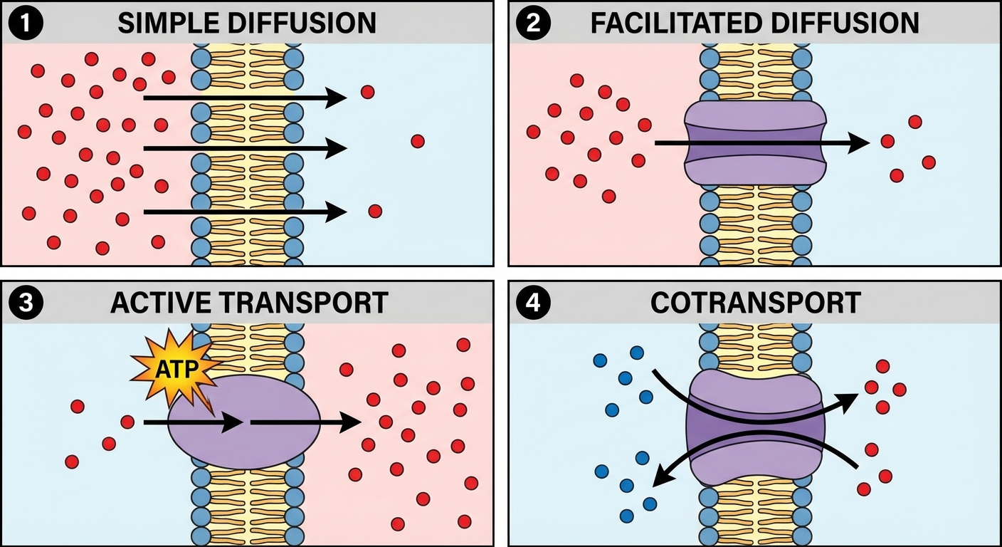
2.6 - 2.9 Transport Mechanisms
Passive Transport
Movement down the concentration gradient (High $\rightarrow$ Low). Requires NO ATP.
- Simple Diffusion: Nonpolar molecules slide through the phospholipids.
- Osmosis: Diffusion of water across a semipermeable membrane.
- Facilitated Diffusion: Movement of polar/charged molecules via transport proteins.
- Channel Proteins: Hydrophilic tunnels (e.g., Aquaporins).
- Carrier Proteins: Change shape to shuttle molecules (e.g., Glucose transport).
Active Transport
Movement against the concentration gradient (Low $\rightarrow$ High). Requires ATP.
- Protein Pumps: Maintain electrochemical gradients.
- $Na^+/K^+$ Pump: Moves 3 $Na^+$ out and 2 $K^+$ in. Essential for nerve transmission.
- Electrogenic Pumps: Generate voltage across membranes (e.g., Proton Pump in plants/fungi).
- Cotransport: Uses the energy from a gradient created by a pump to move a second molecule.
- Example: Sucrose is loaded into plants by coupling it with $H^+$ flowing back into the cell.
Bulk Transport
Used for large macromolecules. Requires ATP.
- Exocytosis: Secretion of vesicles (e.g., neurotransmitters, hormones).
- Endocytosis: Uptake of material.
- Phagocytosis: "Cell Eating" (solids).
- Pinocytosis: "Cell Drinking" (fluids).
- Receptor-Mediated: Highly specific uptake via ligand binding (e.g., cholesterol uptake).
2.8 Osmoregulation & Water Potential
Tip: Water always moves from an area of High Water Potential to Low Water Potential.
The Formula
- $\Psi$ (Psi): Total Water Potential. Pure water in open container = 0.
- $\Psi_p$ (Pressure Potential): Physical pressure exerted by the cell wall.
- Open beaker solution: $\Psi_p = 0$.
- Turgid plant cell: $\Psi_p$ is positive (+).
- $\Psi_s$ (Solute Potential): Also called Osmotic Potential.
- This number is always negative or zero. Adding solute lowers water potential.
- Formula:
The Solute Potential Variables
- $i$: Ionization Constant.
- Sugars (Glucose/Sucrose) do not ionize ($i = 1$).
- Salts (NaCl) break into 2 ions ($i = 2$).
- $C$: Molar Concentration.
- $R$: Pressure Constant ($0.0831$ L bars/mol K).
- $T$: Temperature in Kelvin ($^{\circ}C + 273$).
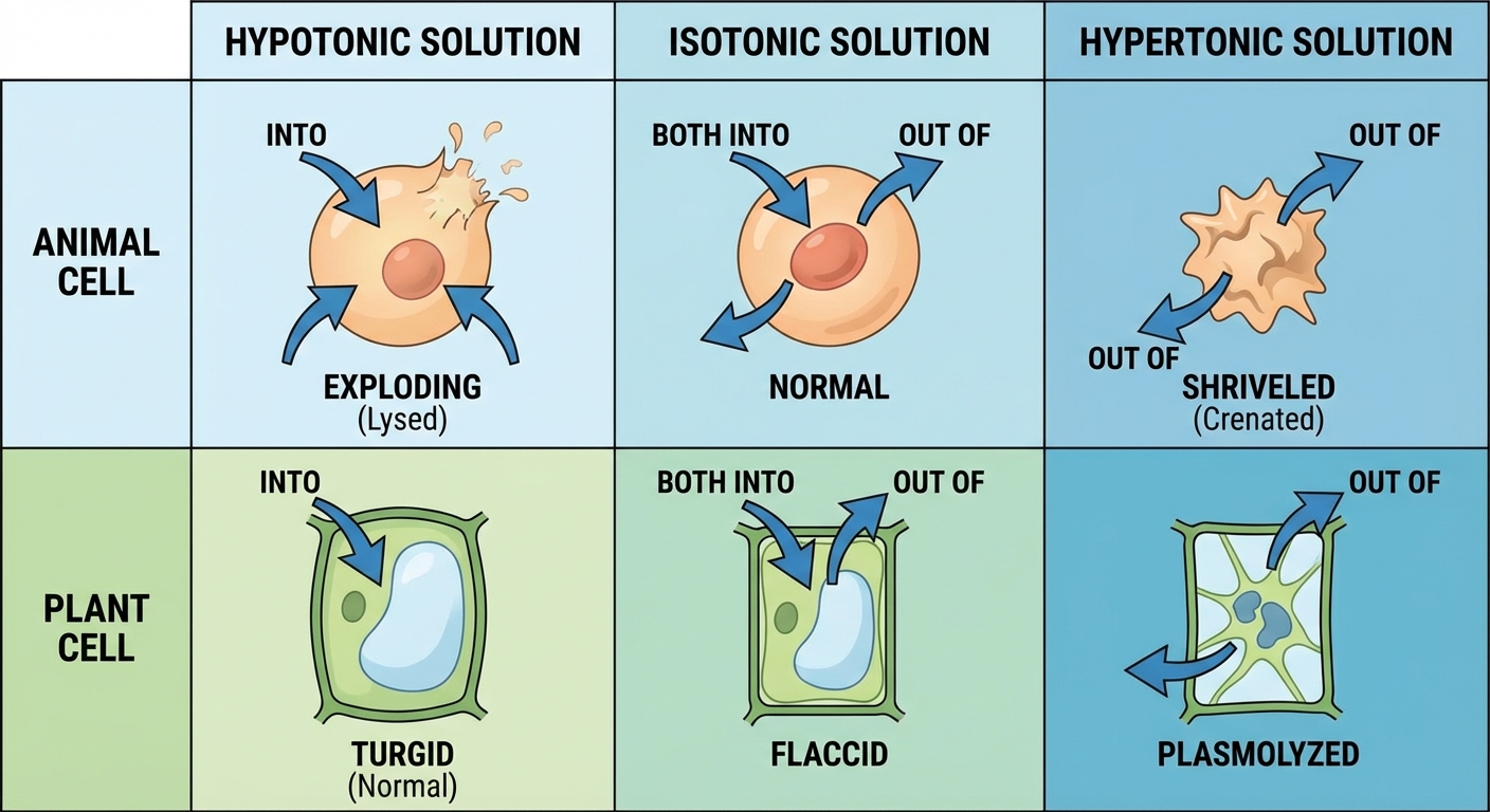
Tonicity Conditions
| Mechanism | Hypotonic Solution (Low Solute) | Isotonic Solution (Equal Solute) | Hypertonic Solution (High Solute) |
|---|---|---|---|
| Water Movement | Enters Cell | Dynamic Equilibrium | Leaves Cell |
| Animal Cell | Lysed (Bursts) | Normal (Optimal) | Shriveled |
| Plant Cell | Turgid (Optimal) | Flaccid | Plasmolyzed |
2.10 & 2.11 Compartmentalization & Evolution
Compartmentalization
By isolating processes, cells gain:
- Efficiency: Enzymes are concentrated with substrates.
- Protection: Lysosomes don't digest the rest of the cell.
Note on Prokaryotes: They lack internal membrane-bound organelles but have internal specialized regions (like nucleoids) and carry out similar functions using their plasma membrane.
Endosymbiotic Theory
Explains how eukaryotes evolved from prokaryotes. It states that an early ancestor engulfed a non-photosynthetic prokaryote (became mitochondria), and later a photosynthetic prokaryote (became chloroplast).
Evidence (Mnemonic: DR. MAD)
- Division: Replicate via binary fission (independent of cell mitosis).
- Ribosomes: Have 70S ribosomes (bacteria-sized).
- Membranes: Double membranes (inner = bacterial, outer = host vesicle).
- Antibiotics: Susceptible to antibiotics targeting bacteria.
- DNA: Have their own circular, naked DNA.
Common Mistakes & Pitfalls
- Sign Errors in Water Potential: Students assume -20 is "larger" than -5. In water potential, $-5 > -20$. Water flows toward the more negative number (towards the high solute).
- Rate vs. Concentration: Facilitated diffusion channels can become saturated (reach a max rate) when all transporters are busy. Simple diffusion does not saturate.
- Active Transport: Just because a protein is involved doesn't mean it's active transport. If it's High $\rightarrow$ Low, it is passive (Facilitated Diffusion). Active transport requires ATP and goes Low $\rightarrow$ High.
- Plant Cell Walls: The cell wall is permeable to small molecules and does not filter like the membrane. What controls entry to the cytoplasm is the plasma membrane inside the wall.
- Hypertonic/Hypotonic: These are relative terms. A solution cannot be "hypotonic" in isolation; it must be hypotonic to the cell.