Mastering Cell Structure and Function: AP Biology Unit 2 Guide
Unit Overview
In AP Biology Unit 2, the central dogma is that structure dictates function. This unit moves beyond simply memorizing the parts of a cell; you must analyze how specific physical attributes (such as the high surface area of a mitochondria's inner membrane) allow for efficient biological processes. This unit provides the physical stage for the metabolic plays (Photosynthesis, Respiration) you will study in Unit 3.
2.1 & 2.2 Subcellular Components
Compartmentalization is the defining characteristic of eukaryotic cells. By using internal membranes, cells create separate local environments (e.g., acidic vs. neutral pH) where specific metabolic reactions can occur without interference.
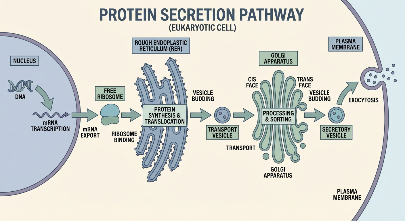
The Protein Assembly Line
Understanding the path of a protein is a frequent exam question. It connects the nucleus, ribosomes, ER, and Golgi.
1. Ribosomes
- Definition: The cellular machinery responsible for translation (protein synthesis). They connect amino acids into polypeptide chains based on mRNA instructions.
- Structure: Consist of two subunits (large and small) made of rRNA (ribosomal RNA) and proteins. They are not membrane-bound.
- Evolutionary Significance: Found in all forms of life (Bacteria, Archaea, Eukarya), serving as strong evidence for common ancestry.
- Locations:
- Free Ribosomes: Float in cytosol; make proteins for intracellular use (e.g., glycolysis enzymes).
- Bound Ribosomes: Attached to the Rough ER; make proteins for secretion, the cell membrane, or lysosomes.
2. Endoplasmic Reticulum (ER)
- Rough ER:
- Studded with ribosomes.
- Function: Synthesizes proteins destined for export or the membrane. It also acts as an intracellular transport canal.
- Smooth ER:
- No ribosomes.
- Function: Synthesizes lipids (phospholipids, steroids), metabolizes carbohydrates, and detoxifies poisons (abundant in liver cells).
3. Golgi Complex
- Structure: Membrane sacs called cisternae.
- Function: The "Post Office." It acts to fold, chemically modify, and package proteins.
- Glycosylation: The Golgi adds carbohydrate tags to proteins, turning them into glycoproteins (vital for cell identification).
- Directionality:
- Cis Face: Receives vesicles from the ER.
- Trans Face: Ships vesicles out to the membrane.
Waste Management & Storage
1. Lysosomes
- Definition: Membrane-enclosed sacs containing hydrolytic enzymes.
- Function: Intracellular digestion.
- Phagocytosis: Digestion of food/bacteria taken in by the cell.
- Autophagy: Recycling the cell's own damaged organelles.
- Apoptosis: Programmed cell death (e.g., webbing between fingers disappearing in a fetus).
- Key Concept: Lysosomal enzymes work best at pH 5. If a lysosome bursts, the neutral cytosol (pH 7) usually deactivates the enzymes, preventing cell self-digestion (unless many burst at once).
2. Vacuoles
- Plants: A large Central Vacuole stores water and ions. It exerts turgor pressure against the cell wall to maintain plant rigidity.
- Protists: Contractile vacuoles pump out excess water to prevent bursting (osmoregulation).
energy Organelles
Both mitochondria and chloroplasts utilize double membranes to compartmentalize energy reactions.
1. Mitochondria
- Function: Site of Cellular Respiration (ATP production).
- Double Membrane Strategy:
- Outer Membrane: Smooth boundary.
- Inner Membrane (Cristae): Highly folded. This folding drastically increases Surface Area, allowing more spaces for the Electron Transport Chain (ETC) and ATP Synthase. More membrane = More ATP.
- Matrix: Fluid center; site of the Krebs Cycle (Citric Acid Cycle).
2. Chloroplasts
- Function: Site of Photosynthesis (in plants/algae).
- Structure:
- Thylakoids: Discs containing chlorophyll; site of Light-Dependent Reactions.
- Grana: Stacks of thylakoids (increase SA).
- Stroma: Fluid surrounding thylakoids; site of the Calvin Cycle (Carbon Fixation).
2.3 Cell Size & Exchange
Biological systems rely on the specific movement of molecules across membranes. This movement is constrained by geometry.
Surface Area-to-Volume Ratio ($SA/V$)
- Volume ($V$): Determines the metabolic needs (how much O$_2$ is needed, how much waste is produced).
- Surface Area ($SA$): Determines the rate of exchange (how fast O$_2$ can enter).
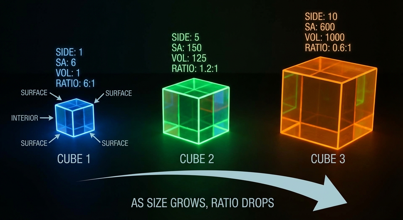
The Problem: As a cell grows, its Volume increases much faster ($s^3$) than its Surface Area ($s^2$).
The Rule: Cells require a high SA/V ratio to survive. Small cells are more efficient at transport than large cells.
Geometric Formulas
For a cube with side $s$:
SA = 6s^2
V = s^3
Ratio = \frac{6s^2}{s^3} = \frac{6}{s}
Structural Adaptations
When cells must be large or specialized for absorption, they modify their shape to increase SA without significantly increasing V:
- Microvilli: Finger-like projections on intestinal lining cells.
- Root Hairs: Extensions on plant roots for water uptake.
- Flattening: Elephant ears (heat dissipation) or fish gills (gas exchange).
2.4 & 2.5 Plasma Membrane Structure
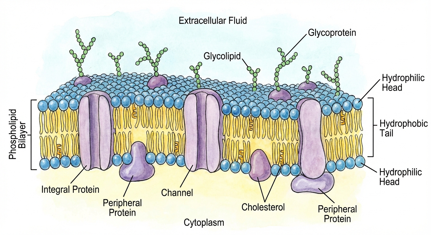
The cell membrane establishes a unique internal environment. It is described by the Fluid Mosaic Model.
1. Phospholipids
The foundation is the bilayer.
- Amphipathic: A molecule with both hydrophilic and hydrophobic regions.
- Head: Phosphate group + Glycerol (Hydrophilic/Polar). Faces the water (inside/outside).
- Tail: Fatty acids (Hydrophobic/Nonpolar). Faces inward, shielding from water.
2. Membrane Components
- proteins:
- Integral (Transmembrane): Span the entire bilayer. Used for transport.
- Peripheral: Loosely bound to the surface (often enzymes or anchor points).
- Cholesterol: A steroid lipid. It acts as a temperature buffer.
- High Temps: Reduces movement (prevents melting).
- Low Temps: Hinders packing (prevents freezing/solidification).
- Carbohydrates: Glycoproteins and Glycolipids function as ID markers (e.g., A/B/O blood types).
3. Selective Permeability
The chemistry of the hydrophobic core dictates what passes:
| Molecule Type | Passage Ability | Examples |
|---|---|---|
| Small, Nonpolar | Pass Freely | $N2$, $O2$, $CO_2$ |
| Small, Polar (uncharged) | Pass Slowly/Minimally | $H_2O$ (mostly needs help) |
| Large, Polar | Blocked (Need Protein) | Glucose, Sucrose |
| Ions (Charged) | Blocked (Need Protein) | $Na^+$, $K^+$, $Cl^-$ |
2.6 - 2.9 Transport Mechanisms
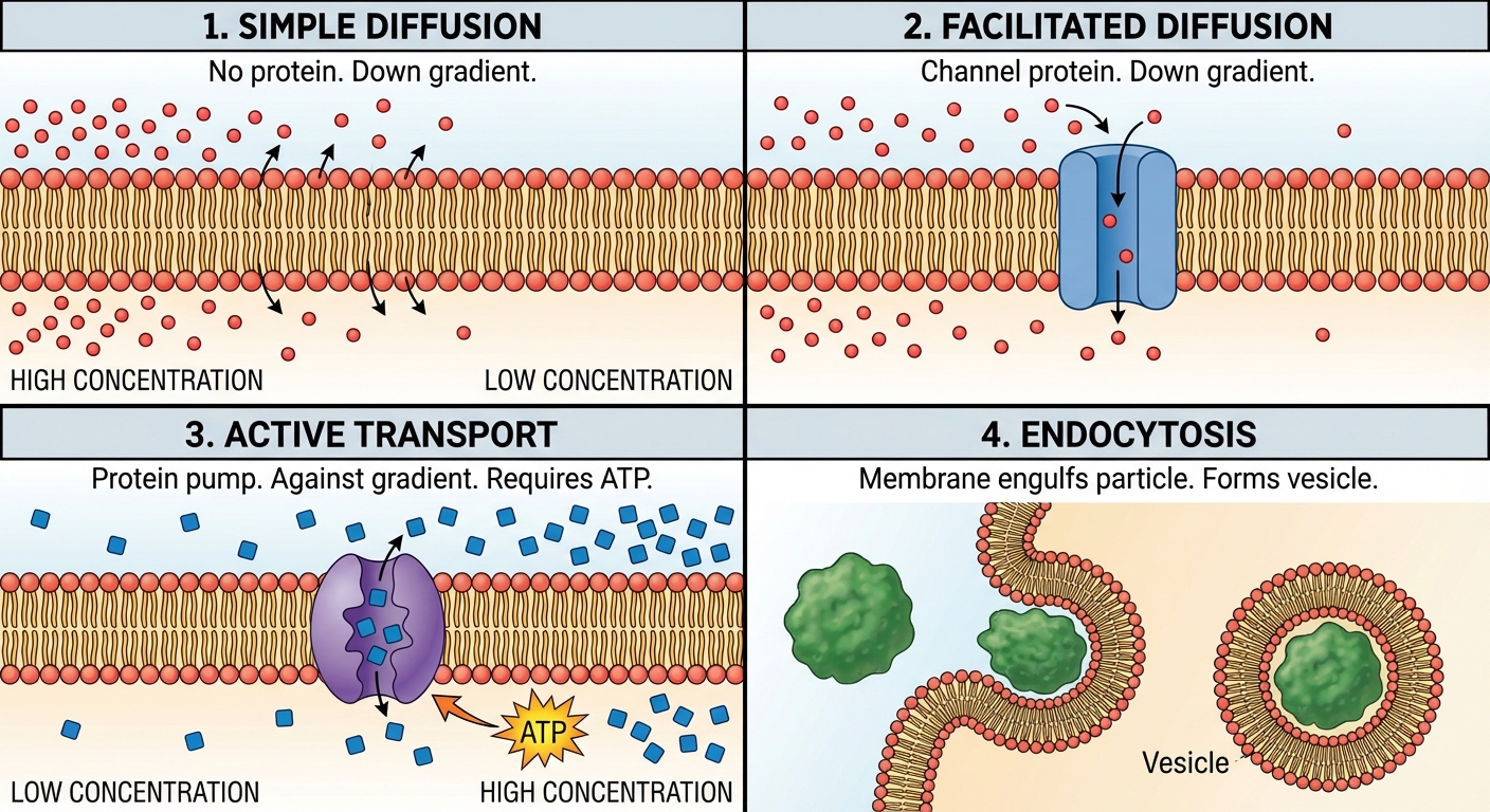
Passive Transport
Movement down the concentration gradient (High $\rightarrow$ Low). Requires NO energy.
- Simple Diffusion: Movement of nonpolar molecules directly through the bilayer.
- Facilitated Diffusion: Movement through transport proteins.
- Channel Proteins: Hydrophilic tunnels (e.g., Aquaporins for water).
- Carrier Proteins: Change shape to shuttle molecules (e.g., Glucose transport).
Active Transport
Movement against the concentration gradient (Low $\rightarrow$ High). Requires Energy (ATP).
- Protein Pumps: Establishment of gradients.
- Na$^+$/K$^+$ ATPase: The most famous pump. Moves 3 $Na^+$ out and 2 $K^+$ in using 1 ATP. This creates an electrochemical gradient essential for nerve function.
- Cotransport: Usage of an electrochemical gradient (created by a pump) to move a second molecule.
- Example: Plants pump $H^+$ out (using ATP). As $H^+$ flows back in (passive), it drags Sucrose with it (active).
Bulk Transport
Used for macromolecules that are too large for proteins. Requires ATP.
- Endocytosis: Taking in.
- Phagocytosis: "Cell Eating" (large solid particles).
- Pinocytosis: "Cell Drinking" (fluid/dissolved solutes).
- Receptor-Mediated: Specific uptake via ligand binding (e.g., LDL cholesterol).
- Exocytosis: Secretion. Vesicles fuse with the plasma membrane to release contents (e.g., neurotransmitters).
2.8 Tonicity and Osmoregulation
Calculations here are guaranteed on the AP Exam.
Osmosis: The diffusion of free water across a selectively permeable membrane. Water always moves from High Water Potential to Low Water Potential.
The Water Potential Formula
\Psi = \Psip + \Psis
- $\Psi$ (Psi): Water Potential. Pure water in an open container = 0.
- $\Psi_p$ (Pressure Potential): Physical pressure.
- Open beaker = 0.
- Turgid plant cell = Positive (+).
- Xylem transport = Negative (tension).
- $\Psi_s$ (Solute Potential): Also called Osmotic Potential.
- Pure water = 0.
- Adding solute makes it Negative.
- Rule: The more solute, the more negative the $\Psi_s$, and the lower the total water potential.
Calculating Solute Potential
\Psi_s = -iCRT
- $i$ (Ionization Constant):
- Sugars (Glucose/Sucrose) do not break apart in water: $i = 1$.
- Salts (NaCl) break into two ions ($Na^+ + Cl^-$): $i = 2$.
- $C$: Molar concentration (Moles/Liter).
- $R$: Pressure constant ($0.0831$ L bars/mol K).
- $T$: Temperature in Kelvin ($^\circ C + 273$).
Tonicity Types
Always compare the environment relative to the cell.
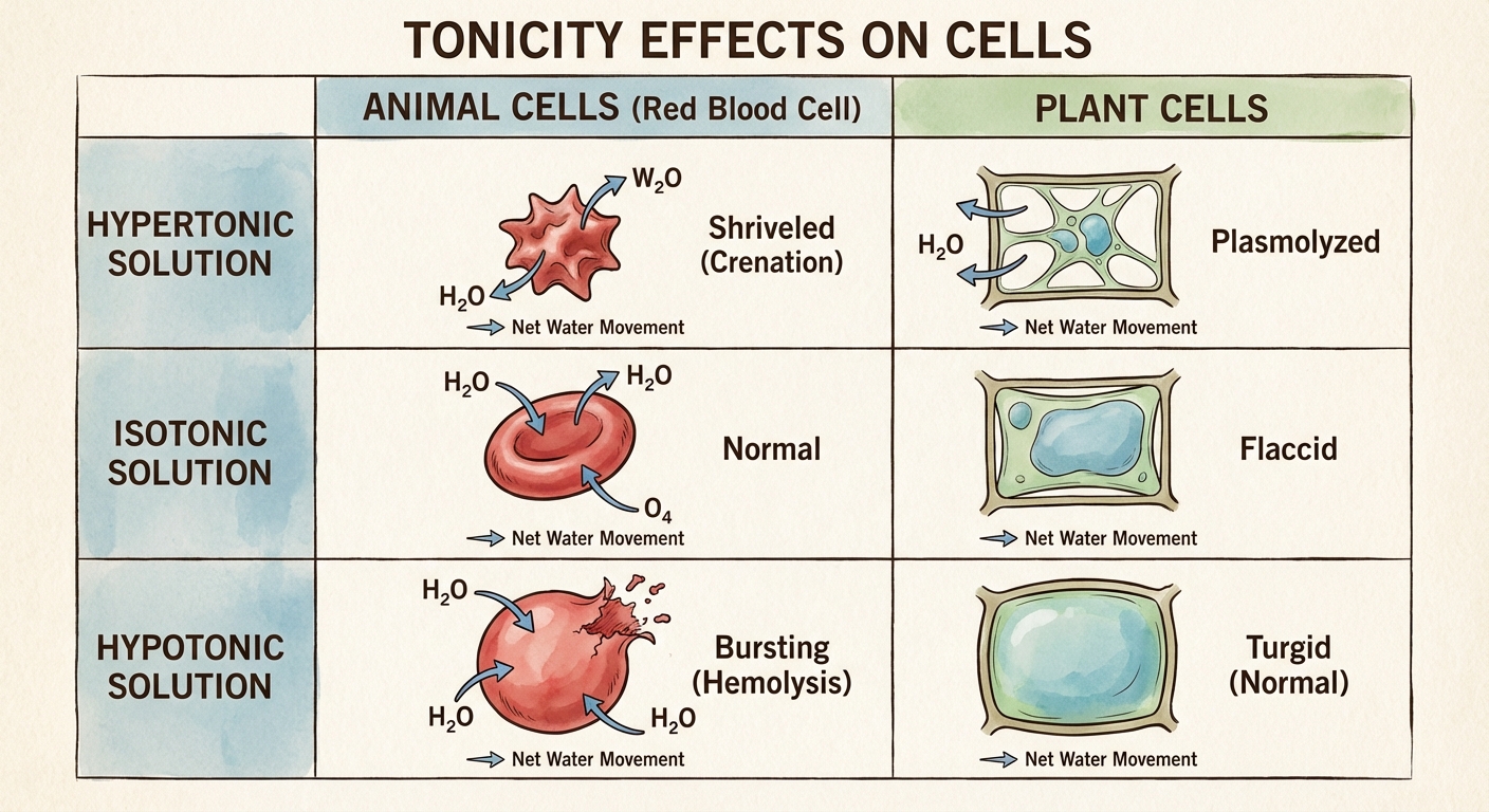
- Hypotonic Environment: Less solute outside, more water outside.
- Water moves INTO the cell.
- Animal Cell: Lysed (Bursts) $\rightarrow$ Bad.
- Plant Cell: Turgid (Firm) $\rightarrow$ Optimal.
- Hypertonic Environment: More solute outside, less water outside.
- Water moves OUT of the cell.
- Animal Cell: Shriveled.
- Plant Cell: Plasmolyzed (membrane pulls away from wall) $\rightarrow$ Deadly.
- Isotonic Environment: Equal solute.
- No net water movement.
- Animal Cell: Normal $\rightarrow$ Optimal.
- Plant Cell: Flaccid (limp).
2.10 & 2.11 Compartmentalization & Evolution
Benefits of Compartmentalization
- Efficiency: Enzymes and substrates are concentrated in one spot.
- Protection: Destructive processes (lysosomes) are contained.
- Gradients: Membranes allow the buildup of gradients (like $H^+$ in mitochondria) required to generate ATP.
Endosymbiotic Theory
How did eukaryotes evolve from prokaryotes?
Theory: An early ancestor of eukaryotic cells engulfed an oxygen-using non-photosynthetic prokaryotic cell. They formed a symbiotic relationship.
- The engulfed cell became the Mitochondrion.
- Later, a cell engulfed a photosynthetic prokaryote, which became the Chloroplast.
Evidence (Mnemonic: DR. MAD)
- D - Division: Mitochondria/Chloroplasts reproduce independently via binary fission, just like bacteria.
- R - Ribosomes: They contain their own ribosomes that are 70S (prokaryotic size), not 80S (eukaryotic size).
- M - Membranes: They have a Double Membrane. The inner is the original bacterial membrane; the outer is the host's vesicle.
- A - Antibiotics: Their protein synthesis is inhibited by antibiotics that target bacteria.
- D - DNA: They have their own circular, naked DNA, distinct from the linear DNA in the nucleus.
Common Mistakes & Pitfalls
- Concentration vs. Electrochemical:
- Mistake: Ignoring the charge.
- Correction: For ions, movement is driven by both the concentration gradient AND the charge gradient (electrochemical). This is why cells expend huge energy on pumps.
- Water Potential Direction:
- Mistake: Thinking water moves to the "more negative" number arbitrarily.
- Correction: Water moves from the mathematically higher number (e.g., -2) to the lower number (e.g., -10). Think of it financially: Water flows towards the debt (solute).
- Active Transport misconception:
- Mistake: Thinking "Protein Channel" = Active Transport.
- Correction: A channel is just a door. If no ATP is used and movement is High $\rightarrow$ Low, it is Facilitated Diffusion (Passive).
- Plants and Mitochondria:
- Mistake: "Plants have chloroplasts, animals have mitochondria."
- Correction: Plants have BOTH. They make sugar in chloroplasts, but they must break it down in mitochondria to get ATP.
- Small vs. Large Cell Efficiency:
- Mistake: "Large cells are better because they hold more."
- Correction: Large cells struggle to feed themselves. High SA/V (small cells) is the metabolic gold standard.