AP Biology Unit 2: Cell Structure and Function
Unit Overview
In AP Biology Unit 2, the core theme is the relationship between structure and function. You must understand not only the components of the cell but how their specific physical attributes (folded membranes, polarity, size) allow them to perform vital processes like protein synthesis, energy capture, and transport. This unit serves as the foundation for Unit 3 (Energetics) and Unit 4 (Cell Communication).
2.1 & 2.2 Subcellular Components
Cells are organized systems where specific organelles facilitate specific metabolic processes. A key concept here is that highly folded membranes increase surface area, allowing for more efficient reaction rates.
The Protein Factory: Ribosomes
Ribosomes are the universal cellular machinery containing rRNA and protein that synthesize proteins according to mRNA sequences. They are found in all forms of life (prokaryotes and eukaryotes), representing common ancestry.
- Structure: Two subunits (large and small) made of ribosomal RNA (rRNA) and proteins. Not membrane-bound.
- Locations & Function:
- Free Ribosomes: Floating in the cytosol. They generally synthesize proteins functioning within the cytosol (e.g., enzymes for glycolysis).
- Bound Ribosomes: Attached to the Rough ER. They synthesize proteins usually destined for secretion, the cell membrane, or lysosomes.
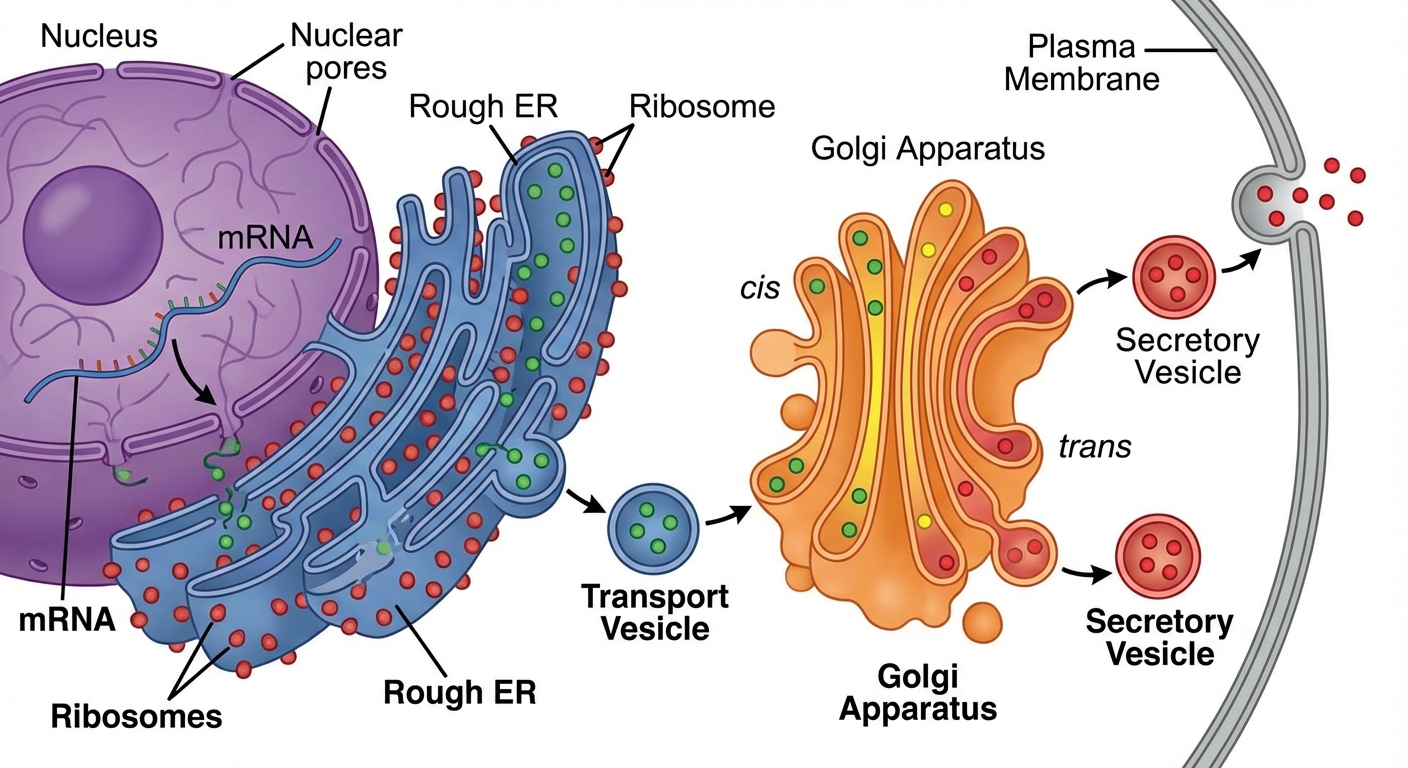
The Endomembrane System
This group of organelles works together to modify, package, and ship proteins and lipids.
1. Endoplasmic Reticulum (ER)
- Rough ER: Studded with ribosomes. Compartmentalizes the cell and provides mechanical support. Main function: protein synthesis for export.
- Smooth ER: No ribosomes. Functions include lipid synthesis, detoxification (abundant in liver cells), and carbohydrate metabolism.
2. The Golgi Complex
- Structure: Flattened membrane sacs called cisternae.
- Function: Correct folding and chemical modification of newly synthesized proteins (e.g., glycosylation—adding sugar tags) and packaging for protein trafficking.
- Cis face: Receives vesicles from the ER.
- Trans face: Ships vesicles to the plasma membrane or other organelles.
3. Lysosomes
- Structure: Membrane-enclosed sacs containing hydrolytic enzymes.
- Function: Intracellular digestion.
- Autophagy: Recycling the cell’s own organic materials/damaged organelles.
- Apoptosis: Programmed cell death.
- Important: Hydrolytic enzymes function best in acidic environments (pH ~5). The lysosome maintains this pH distinct from the neutral cytosol.
4. Vacuoles
- Structure: Large membrane-bound sacs.
- Function: Storage of water, macromolecules, and waste.
- Plants: Have a large Central Vacuole that aids in retention of water for turgor pressure.
- Protists: Some have contractile vacuoles for osmoregulation (pumping out water).
Energy-Capturing Organelles
Both mitochondria and chloroplasts specialize in energy transformation through double-membrane systems.
1. Mitochondria
- Function: Site of Cellular Respiration (ATP production).
- Outer Membrane: Smooth.
- Inner Membrane: Highly folded into cristae. This folding maximizes surface area for the Electron Transport Chain (ETC).
- Matrix: The fluid-filled center where the Krebs Cycle occurs.
2. Chloroplasts
- Function: Site of Photosynthesis (in plants/algae).
- Thylakoids: Membranous sacs stacked into grana. The abundance of thylakoid membranes maximizes the surface area for the light-dependent reactions.
- Stroma: The fluid surrounding thylakoids; site of the Calvin Cycle (carbon fixation).
| Feature | Mitochondria | Chloroplasts |
|---|---|---|
| Function | ATP Generation (Respiration) | Sugar Generation (Photosynthesis) |
| Key Structure | Cristae (folded inner membrane) | Thylakoids (stacked sacs) |
| Location | Almost all Eukaryotes | Plants & Algae |
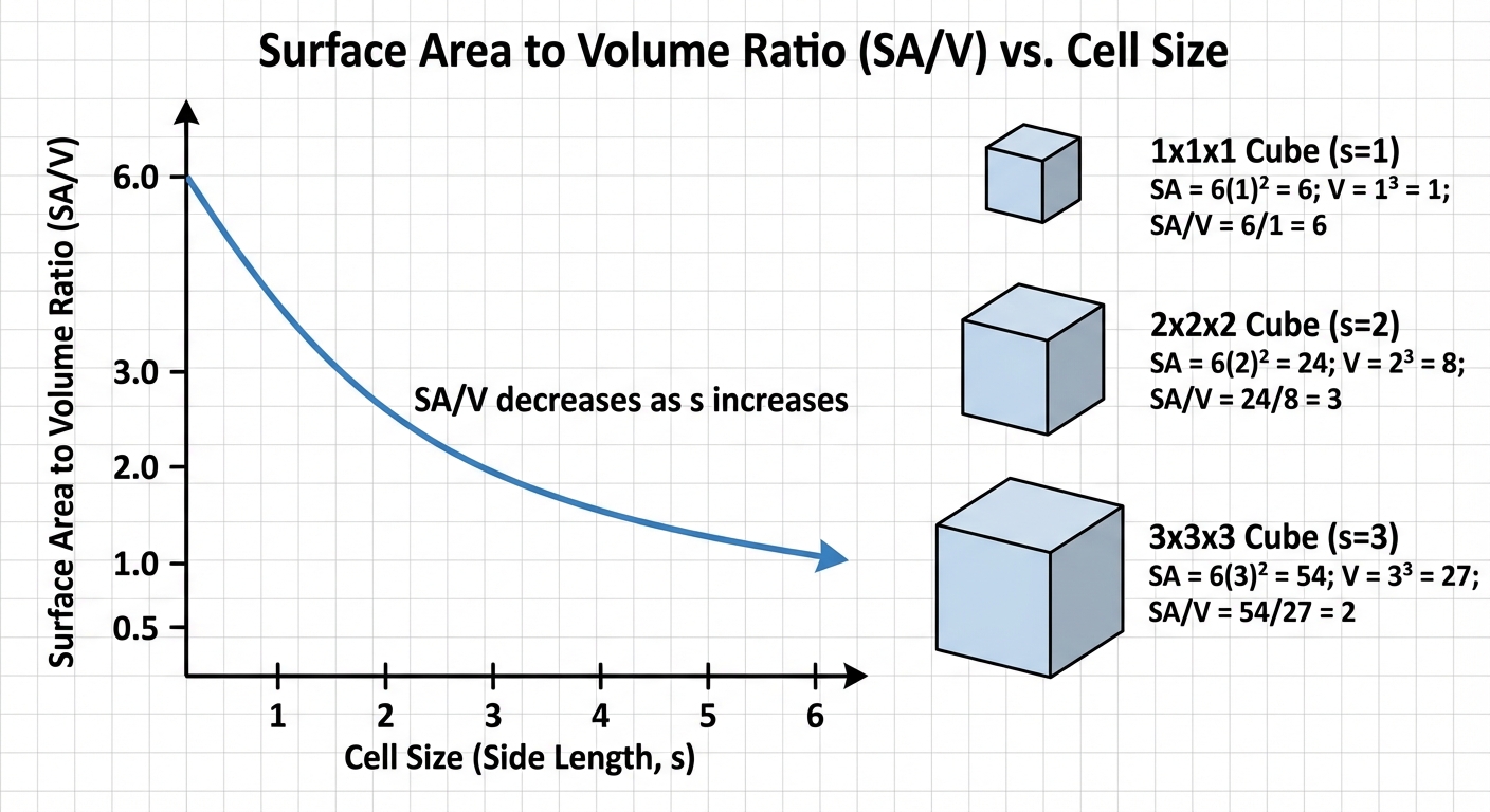
2.3 Cell Size
Cells are limited in size by the physics of diffusion. The movement of materials into (nutrients) and out of (waste) the cell relies on the Surface Area-to-Volume Ratio ($SA/V$).
The Math of SA/V
As a cell gets larger, its volume increases much faster than its surface area.
- Volume ($s^3$): Determines metabolic demand (how much food allows/waste produced).
- Surface Area ($s^2$): Determines the rate of exchange.
SA{cube} = 6s^2 V{cube} = s^3
Conclusion: Smaller cells have a higher $SA/V$ ratio, making them more efficient at exchanging materials with the environment.
Adaptations for Efficiency
Cells that must be large or specialized for absorption utilize structural adaptations to increase surface area:
- Root Hairs: Increase absorption of water in plants.
- Microvilli: Finger-like projections on intestinal cells.
- Flattened Shape: Decreases distance for diffusion (e.g., Red blood cells, elephant ears for heat dissipation).
2.4 & 2.5 Plasma Membrane Structure & Permeability
The cell membrane establishes an internal environment that is different from the external environment.
The Fluid Mosaic Model
Moving membranes composed of a phospholipid bilayer with embedded proteins, steroids, and carbohydrates.
- Phospholipids: Amphipathic molecules.
- Hydrophilic Head: Phosphate group; faces aqueous environments (inside/outside).
- Hydrophobic Tail: Fatty acids; faces inward, away from water.
- Membrane Proteins:
- Integral Proteins: Span the membrane (transmembrane).
- Peripheral Proteins: Loosely bound to the surface.
- Cholesterol: Regulates membrane fluidity. At high temps, it stabilizes the membrane; at low temps, it prevents tight packing (freezing).
Selective Permeability
The structure of the phospholipids determines what can pass directly through the bilayer:
- Pass Freely: Small, nonpolar molecules ($\text{N}2$, $\text{O}2$, $\text{CO}_2$).
- Need Help (Transport Proteins): Large polar molecules (Glucose), ions ($\text{Na}^+, \text{K}^+$), and water (polar).
Cell Walls: Structural boundaries in plants (cellulose), fungi (chitin), and bacteria (peptidoglycan). They provide shape and prevent cellular rupture, but are generally permeable to most substances unlike the membrane.
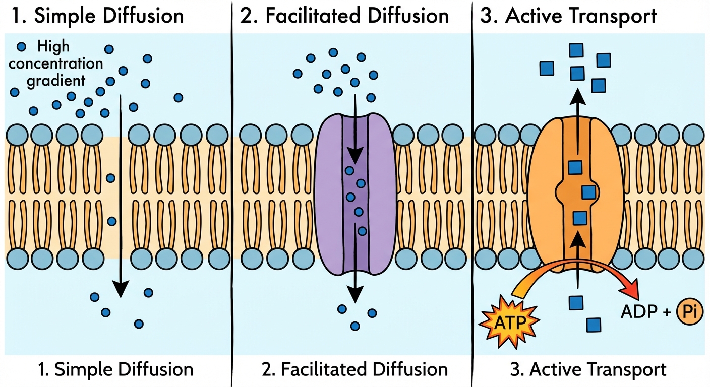
2.6 - 2.9 Membrane Transport
Passive Transport
Movement without metabolic energy (ATP). Molecules move from High Concentration $\rightarrow$ Low Concentration.
- Simple Diffusion: Nonpolar molecules slip through the phospholipid bilayer.
- Facilitated Diffusion: Movement of molecules through transport proteins (channels or carriers).
- Example: Aquaporins are channels specifically for water.
- Example: Ion channels (gated) for $\text{Na}^+$ or $\text{K}^+$.
Active Transport
Requires metabolic energy (usually ATP) to move molecules against the gradient (Low $\rightarrow$ High).
- Pumps: e.g., The Sodium-Potassium Pump ($\text{Na}^+/\text{K}^+$ ATPase). Maintains membrane potential.
- Cotransport: Uses the energy from an electrochemical gradient (generated by a pump) to transport a different ion/molecule.
Bulk Transport (Active)
- Endocytosis: Cell takes in material by forming vesicles from the plasma membrane.
- Phagocytosis: Cell eating.
- Pinocytosis: Cell drinking.
- Exocytosis: Internal vesicles fuse with the membrane to secrete large macromolecules (e.g., neurotransmitters, proteins).
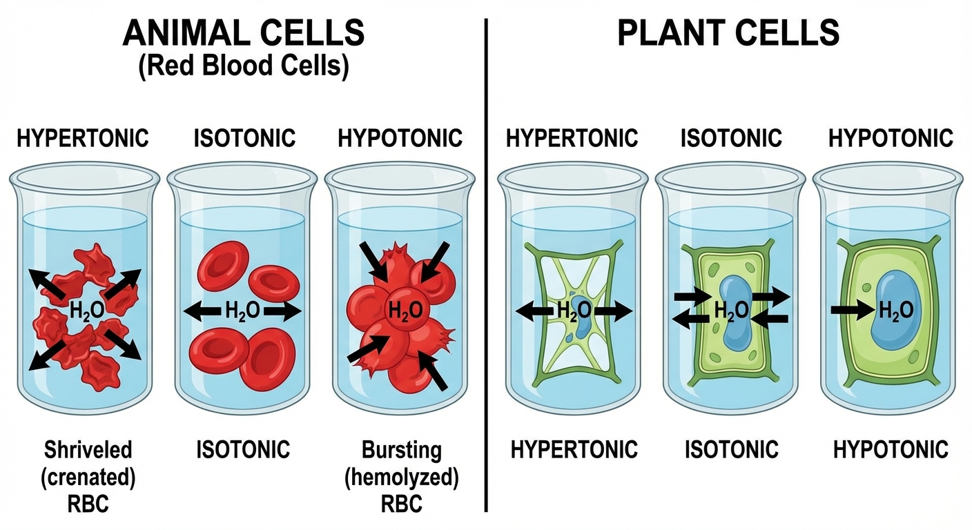
2.8 Tonicity and Osmoregulation
Water moves by osmosis from areas of High Water Potential to Low Water Potential (or Low Solute $\rightarrow$ High Solute).
Water Potential Formula
This is a guaranteed math question on the AP exam.
\Psi = \Psip + \Psis
- $\Psi$ (Psi): Total water potential.
- $\Psi_p$: Pressure potential. Open containers = 0. Turgor pressure in plants > 0.
- $\Psi_s$: Solute potential. Always $\le 0$. Adding solute lowers water potential.
Calculating Solute Potential:
\Psi_s = -iCRT
- $i$: Ionization constant (Sucrose/sugar = 1, NaCl = 2).
- $C$: Molar concentration.
- $R$: Pressure constant (usually 0.0831).
- $T$: Temperature in Kelvin ($^\circ\text{C} + 273$).
Tonicity Conditions
| Condition | Definition | Effect on Animal Cell | Effect on Plant Cell |
|---|---|---|---|
| Hypotonic | Environ. has less solute (more water) | Lysed (Bursts) | Turgid (Normal) - Wall protects it |
| Isotonic | Equal solute levels | Normal | Flaccid (limp) |
| Hypertonic | Environ. has more solute (less water) | Shriveled | Plasmolyzed (membrane pulls away from wall) |
2.10 & 2.11 Compartmentalization & Origins
Compartmentalization separates different metabolic processes within the same cell using membranes.
Benefits
- Efficiency: Enzymes/substrates are concentrated in specific areas.
- Protection: Hydrolytic enzymes in lysosomes don't destroy the rest of the cell.
- Membrane Surface Area: Folding (mitochondria/chloroplasts) allows for more ATP production.
The Endosymbiotic Theory
Explains the origin of eukaryotic cells. Mitochondria and chloroplasts were once free-living prokaryotes engulfed by an ancestral cell.
Evidence (The "DR. D" Mnemonic):
- D - DNA: They have their own circular, naked DNA.
- R - Ribosomes: They have prokaryotic-sized (70S) ribosomes.
- D - Double Membrane: Inner membrane reflects the original bacterium; outer membrane reflects the host's vesicle.
- Reproduction: They replicate independently via binary fission.
Common Mistakes & Pitfalls
- Protein Path Trajectory:
- Mistake: Thinking proteins go from Smooth ER to Golgi.
- Correction: The Correct path for secreted proteins is: Ribosome $\rightarrow$ Rough ER $\rightarrow$ Transport Vesicle $\rightarrow$ Golgi $\rightarrow$ Secretory Vesicle $\rightarrow$ Plasma Membrane.
- Water Potential Direction:
- Mistake: Thinking water moves to high water potential.
- Correction: Water flows High $\Psi$ $\rightarrow$ Low $\Psi$. (Think of it like a ball rolling down a hill).
- Hypertonic vs. Hypotonic:
- Mistake: Confusing the terms. "Hyper" means High/Above (high solute).
- Correction: If the beaker is Hypertonic, water leaves the cell (cell shrinks). If the beaker is Hypotonic, water enters the cell (cell swells/pops).
- Plant Mitochondria:
- Mistake: Thinking plants only have chloroplasts.
- Correction: Plants have both. Chloroplasts make the sugar; Mitochondria break it down for ATP.
- Passive vs. Active:
- Mistake: Thinking all protein channels are active transport.
- Correction: Facilitated diffusion uses protein channels but requires no energy because it goes down the concentration gradient.