AP Biology Unit 4: Signal Transduction and the Cell Cycle
Cell Communication Mechanisms (Topics 4.1 - 4.2)
Overview of Cellular Signaling
Cells must interact with their environment and other cells to maintain homeostasis and coordinate function.
Evolutionary Significance
Signal transduction pathways are highly conserved across evolutionary history. The similarities in pathways between unicellular organisms (like yeast mating factors) and complex multicellular mammals suggest that these mechanisms evolved in a common ancestor long ago.
Modes of Cell Communication
Cells communicate via chemical signals either through direct contact or over distances.
| Type | Description | Key Examples |
|---|---|---|
| Juxtacrine (Direct Contact) | Physical contact between cells or cell-to-cell channels. | Plasmodesmata (plants), Gap Junctions (animals), Immune cell interaction (Antigen Presenting Cells). |
| Paracrine (Short Distance) | Signals released into the extracellular fluid to affect nearby target cells. | Neurotransmitters moving across a synapse; Morphogens in embryonic development. |
| Autocrine (Self Signaling) | A cell secretes a signal that binds to receptors on its own surface. | T-cell activation in the immune system; Cancer cells triggering their own growth. |
| Endocrine (Long Distance) | Hormones travel through the circulatory system (blood) to reach distant target cells. | Insulin, Glucagon, Human Growth Hormone (HGH). |
Note on Taxis: While often discussed in ecology, unicellular organisms exhibit taxis (movement toward/away from stimulus).
- Chemotaxis: Bacterial movement toward food (positive) or away from toxins (negative).
- Quorum Sensing: Bacteria use chemical signals to detect population density and coordinate group behaviors (e.g., biofilm formation).
The Three Stages of Signaling (Topic 4.3)
Signal Transduction is the process by which an external signal is converted into a functional cellular response. It occurs in three defined steps:
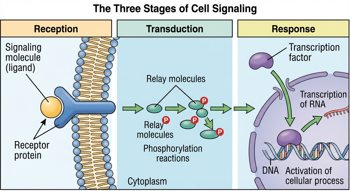
1. Reception
Detection of a signaling molecule coming from outside the cell.
- The Ligand: A specific signaling molecule that binds to a receptor.
- The Concept: The binding is highly specific (Lock and Key model). When a ligand binds, it causes a conformational shape change in the receptor protein. This shape change is the initial "transduction" of the signal.
Types of Receptors:
- G-Protein Coupled Receptors (GPCRs):
- The largest family of cell-surface receptors.
- Mechanism: Ligand binds $\rightarrow$ GPCR changes shape $\rightarrow$ GPCR binds to an intracellular G-protein $\rightarrow$ G-protein swaps GDP (inactive) for GTP (active) $\rightarrow$ G-protein triggers the next step.
- Ligand-Gated Ion Channels:
- Act as a "gate" for ions.
- Mechanism: Ligand binds $\rightarrow$ Channel opens $\rightarrow$ Ions ($Na^+$, $Ca^{2+}$) flow through down their concentration gradient $\rightarrow$ Rapid cellular response (essential for nervous system signaling).
- Intracellular Receptors:
- Found in the cytoplasm or nucleus.
- Ligands must be hydrophobic (nonpolar) or very small (e.g., Steroid hormones like Testosterone or Nitric Oxide) to pass through the phospholipid bilayer unaided.
- These often act as transcription factors, turning genes on or off directly.
2. Transduction
converts the signal to a form that can bring about a specific cellular response. This acts as a relay race.
- Signal Amplification: A single ligand can activate millions of molecules. One receptor activates many G-proteins, which activate many enzymes, etc.
- Phosphorylation Cascades: A series of sequential phosphorylation reactions.
- Protein Kinase: An enzyme that transfers phosphate groups from ATP to a protein (activates the protein).
- Protein Phosphatase: An enzyme that removes phosphate groups from proteins (deactivates the protein/shuts down the pathway).
- Second Messengers: Small, non-protein, water-soluble molecules that spread the signal throughout the cytoplasm.
- Cyclic AMP (cAMP): Created from ATP by the enzyme Adenylyl Cyclase.
- Calcium Ions ($Ca^{2+}$): Used extensively in muscle contraction and secretion.
3. Response
The specific cellular activity triggered by the transducer signal.
- Nuclear Response: Turning genes on/off (Transcription factors modulating protein synthesis).
- Cytoplasmic Response: Activating an existing enzyme or opening/closing an ion channel.
- Apoptosis: Programmed cell death (e.g., forming fingers during development by killing the cells between them).
Changes in Signal Transduction (Topic 4.4)
Alterations in the pathway can lead to disease or altered responses.
- Mutations: A mutation in the reef receptor gene may create a receptor that signals even without a ligand (constitutively active), potentially leading to cancer.
- Inhibitors/Blockers: Chemicals (poisons or drugs) can bind to receptors to block the ligand (e.g., Antihistamines block histamine receptors).
Feedback Mechanisms (Topic 4.5)
Living systems use feedback loops to maintain their internal environments (Homeostasis) or to drive processes to completion.
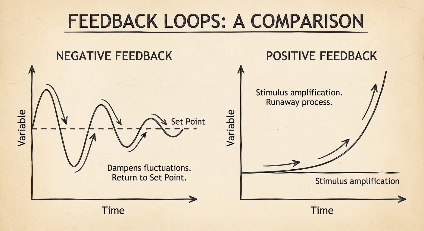
Negative Feedback
Mechanisms that return a system to a set point. The output reduces the original effect of the stimulus.
- Goal: Stability and Homeostasis.
- Example: Blood Sugar Regulation
- High Glucose $\rightarrow$ Pancreas secretes Insulin $\rightarrow$ Cells take up glucose $\rightarrow$ Levels drop.
- Low Glucose $\rightarrow$ Pancreas secretes Glucagon $\rightarrow$ Liver releases glucose $\rightarrow$ Levels rise.
Positive Feedback
Mechanisms that amplify physiological changes. The output increases the original stimulus, moving the system away from the set point.
- Goal: Drive a process to completion/climax.
- Example: Childbirth
- Head pushes on cervix $\rightarrow$ Nerve impulses $\rightarrow$ Pituitary releases Oxytocin $\rightarrow$ Uterus contracts $\rightarrow$ Head pushes harder $\rightarrow$ More Oxytocin (Cycle repeats until birth).
- Example: Apple Ripening
- Ripe apple releases Ethylene gas $\rightarrow$ Signals neighbors to ripen $\rightarrow$ Neighbors release more Ethylene.
The Cell Cycle (Topic 4.6)
The cell cycle is the life of a cell from formation to its own division. It ensures the genome is passed faithfully to offspring.
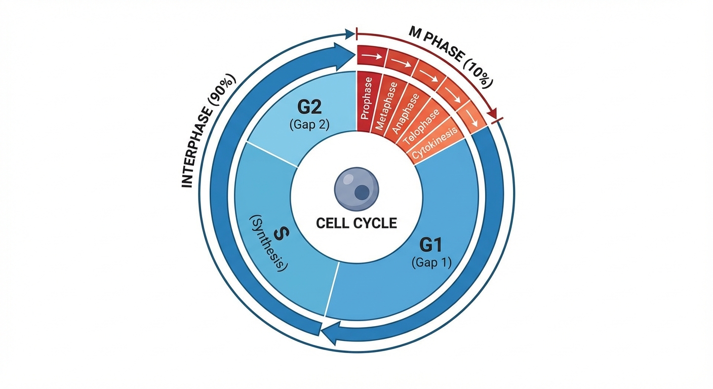
Interphase (90% of the Cycle)
Interphase is NOT a resting phase; the cell is metabolically active.
- G1 Phase (First Gap): The cell grows, produces organelles, and carries out normal functions.
- G0 Phase: A non-dividing state. Some cells (neurons, muscle cells) exit the cycle and stay here permanently. Others (liver cells) can be called back from G0.
- S Phase (Synthesis): DNA Replication occurs here. Chromosomes are duplicated.
- Terminology: Before S phase, a chromosome is 1 strand. After S phase, a chromosome consists of two identical sister chromatids connected at the centromere.
- G2 Phase (Second Gap): The cell continues to grow and prepares for division (producing microtubules, etc.).
M-Phase: Mitosis and Cytokinesis
Mitosis is the division of the nucleus. Cytokinesis is the division of the cytoplasm.
Stages of Mitosis (PMAT)
- Prophase:
- Chromatin condenses into visible chromosomes.
- Nucleoli disappear; Nuclear envelope breaks down.
- Mitotic spindle begins to form.
- Metaphase:
- Chromosomes align at the Metaphase Plate (equator).
- Spindle fibers (microtubules) attach to the Kinetochores at the centromeres.
- Anaphase:
- Cohesin proteins holding sister chromatids together cleave.
- Sister chromatids separate and move to opposite poles.
- Crucial: Once separated, they are considered individual chromosomes.
- Telophase:
- Two daughter nuclei form.
- Chromosomes decondense into chromatin.
Cytokinesis
- Animal Cells: A cleavage furrow forms (contractile ring of microfilaments) pinching the cell in two.
- Plant Cells: A cell plate forms from Golgi vesicles, eventually creating a new cell wall.
Regulation of the Cell Cycle (Topic 4.7)
Cell Cycle Checkpoints
Control points where stop/go-ahead signals regulate the cycle.
- G1 Checkpoint (Restriction Point): The most important. Checks for cell size, nutrients, and DNA damage. If denied, cell enters G0.
- G2 Checkpoint: Checks if DNA replication was successful and undamaged.
- M Checkpoint (Spindle Checkpoint): Occurs during Metaphase. Ensures all chromosomes are attached to spindle fibers. If not, Anaphase is blocked (preventing incorrect chromosome numbers).
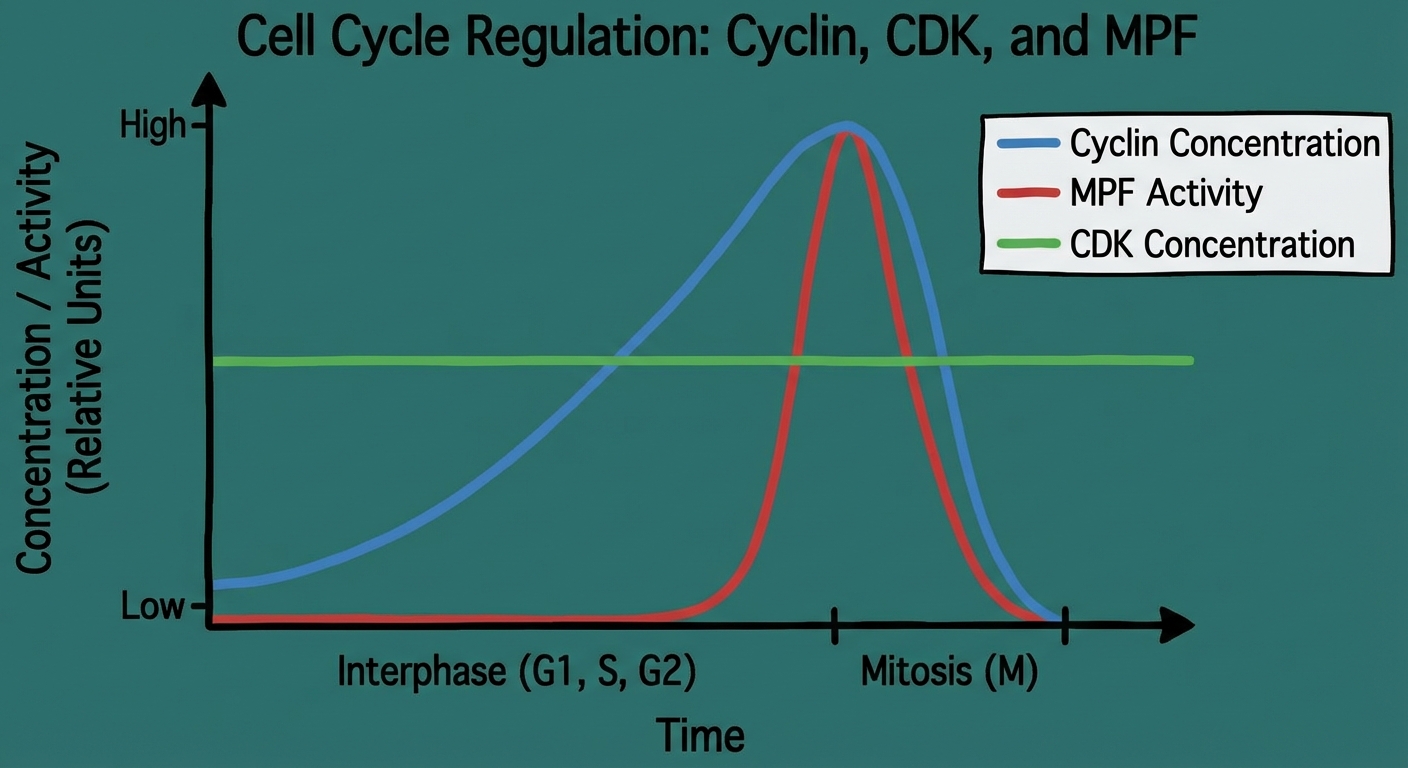
Molecular Control System
The cell cycle is driven by specific signaling molecules present in the cytoplasm.
- Cyclins: Proteins whose concentration fluctuates (cycles) throughout the cell cycle.
- Cyclin-Dependent Kinases (CDKs): Enzymes that are constant in concentration but inactive unless attached to a cyclin.
- MPF (Maturation-Promoting Factor): A Cyclin-CDK complex that triggers the cell's passage into the M phase.
- Mechanism: Cyclin synthesis rises in S and G2 $\rightarrow$ binds to CDK $\rightarrow$ forms MPF $\rightarrow$ Mitosis begins $\rightarrow$ Cyclin degrades during Anaphase $\rightarrow$ CDK is recycled.
Cancer: Loss of Cell Cycle Control
Cancer arises when cells ignore checkpoints and divide uncontrollably.
- Transformation: Conversion of a normal cell to a cancerous one.
- Tumors: Masses of abnormal cells.
- Benign: Stay at the original site.
- Malignant: Invade surrounding tissues (Invasive).
- Metastasis: Spread of cancer cells to distant locations via blood or lymph.
Genetic Basis of Cancer:
- Proto-oncogenes: Normal genes that stimulate cell growth. If mutated into Oncogenes, they become hyperactive (like a gas pedal stuck down).
- Tumor-Suppressor Genes: Normal genes that inhibit cell division and repair DNA (e.g., p53). If mutated/defective, the "brakes" fail.
- The Job of p53: If DNA damage is detected, p53 halts the cycle for repair. If repair is impossible, p53 triggers apoptosis.
Common Mistakes & Pitfalls
- Growth vs. Division: Students often think Interphase is "resting." Correct: It is a period of intense growth and metabolic activity.
- Chromosomes vs. Chromatids:
- 1 Chromosome (G1) = 1 DNA molecule.
- 1 Chromosome (G2/Metaphase) = 2 Sister Chromatids (but still counts as 1 chromosome unit until they separate).
- Tip: Count the Centromeres to count the Chromosomes.
- Positive Feedback: Often confused with "good" outcomes. Positive feedback merely means amplification. It can be fatal (e.g., overheating/fever loops) if not broken.
- Kinase vs. Phosphatase: Remember Kinase adds energy (ATP/Phosphorus) giving the protein a "Kick" to start working.
- Binary Fission vs. Mitosis: Prokaryotes (Bacteria) do binary fission. Eukaryotes do Mitosis. Do not mix them up.